Demographics:
54 years old, Female
Indication:
Headache
Findings
- Large, peripherally-enhancing, centrally necrotic mass bridging the right frontal and parietal lobes measuring 4.5 x 4.2 x 4.5 cm with extensive T2/FLAIR signal hyperintensity in the surrounding white matter
- Restricted diffusion correlating with the enhancing components
- Susceptibility artifact correlating with the area of central necrosis
- Additionally masslike area of restricted diffusion in the posterior left temporal lobe with a corresponding focus of enhancement along its anterior margin
- Mass effect in the right cerebral hemisphere resulting in local sulcal effacement, crowding of the right lateral ventricle, and 9 mm right-to-left midline shift
- Mild dilation of the temporal horn of the right lateral ventricle concerning for entrapment
Annotated Images & Illustrations
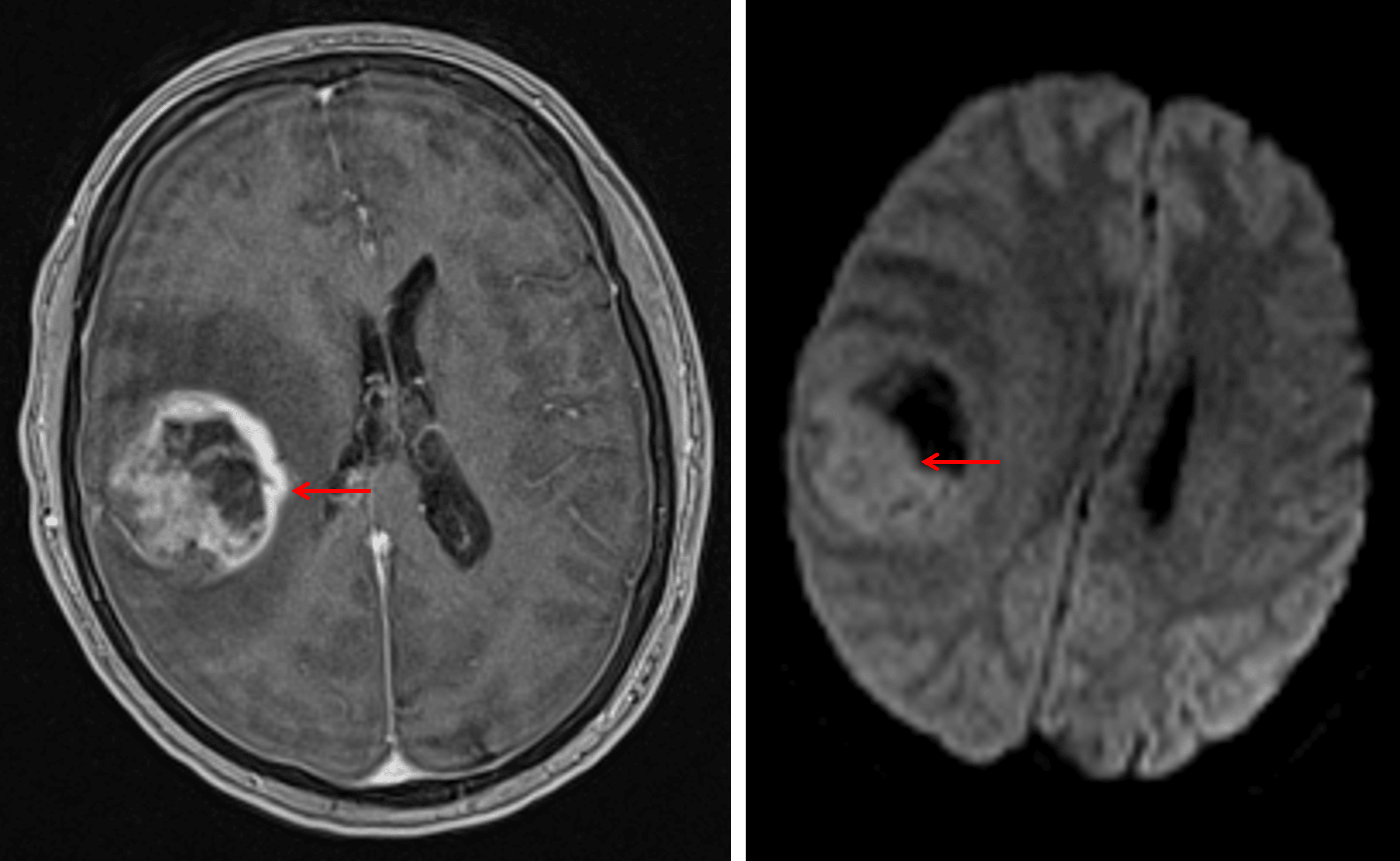
Heterogeneously enhancing mass with reduced diffusivity corresponding with the enhancing components (red arrows), most suspicious for a high-grade glial neoplasm.
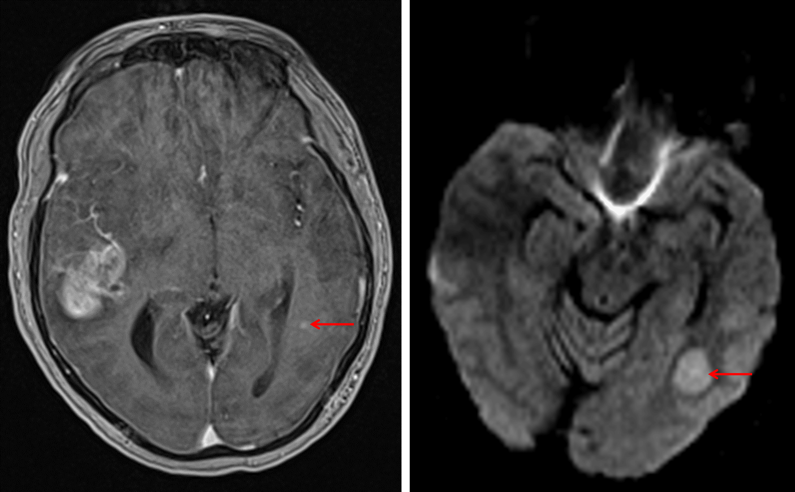
Additional masslike area of restricted diffusion in the posterior left temporal lobe with a nodular focus of enhancement along its anterior margin (red arrows).
Diagnosis
Gliosarcoma
Key Imaging Features
Become a PRO member to unlock the key imaging features
Differential Diagnosis
Become a PRO member to unlock the differential diagnosis
Discussion
Pearls
Become a PRO member to unlock the pearls