Demographics:
6 years old, Female
Indication:
Headache
Findings
CT
- Masslike area of hypoattenuation involving the dorsal pons and middle cerebellar peduncles with mass effect on the fourth ventricle
- Mucosal thickening and fluid in the bilateral sphenoid sinuses and posterior ethmoid air cells
MRI
- Masslike area of T2/FLAIR signal hyperintensity involving the pons, midbrain including the cerebral peduncles, left eccentric medulla, and left greater than right middle cerebellar peduncles
- Area of relative restricted diffusion in the ventral aspect of the pons
- The mass bulges into the prepontine cistern and at least partially encases the basilar artery
- Associated mass effect on the fourth ventricle without overt hydrocephalus
- No corresponding enhancement
Annotated Images & Illustrations
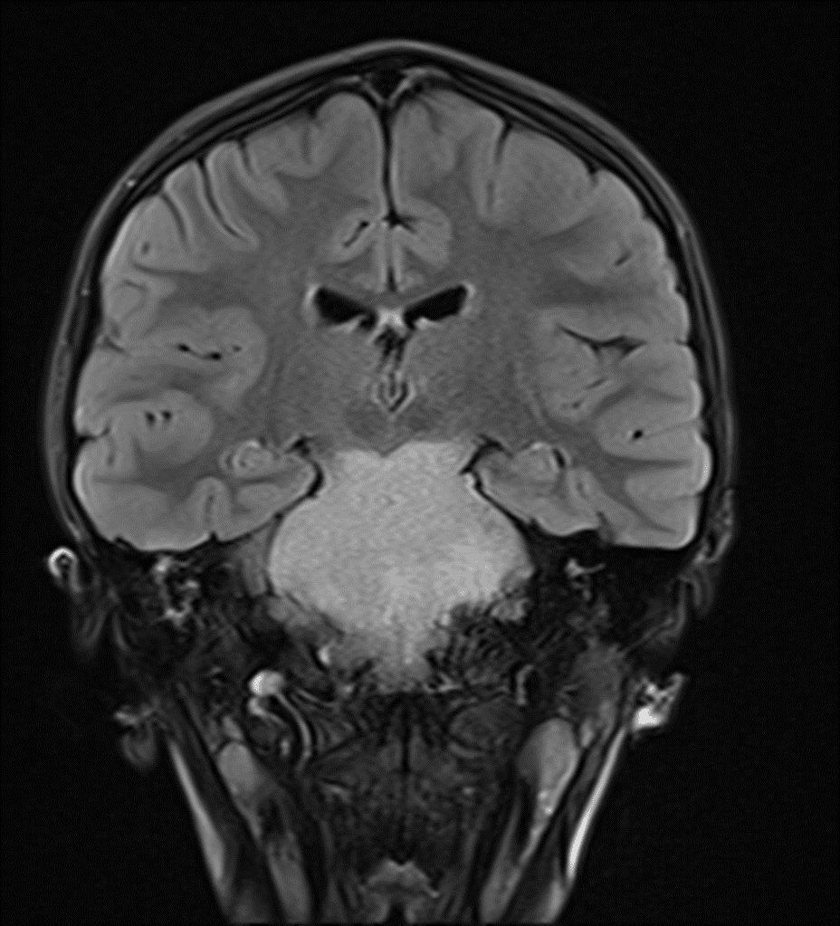
Homogeneous FLAIR hyperintense mass involving the entire pons and extending into the midbrain and medulla, which is a classic imaging appearance for a diffuse midline glioma.
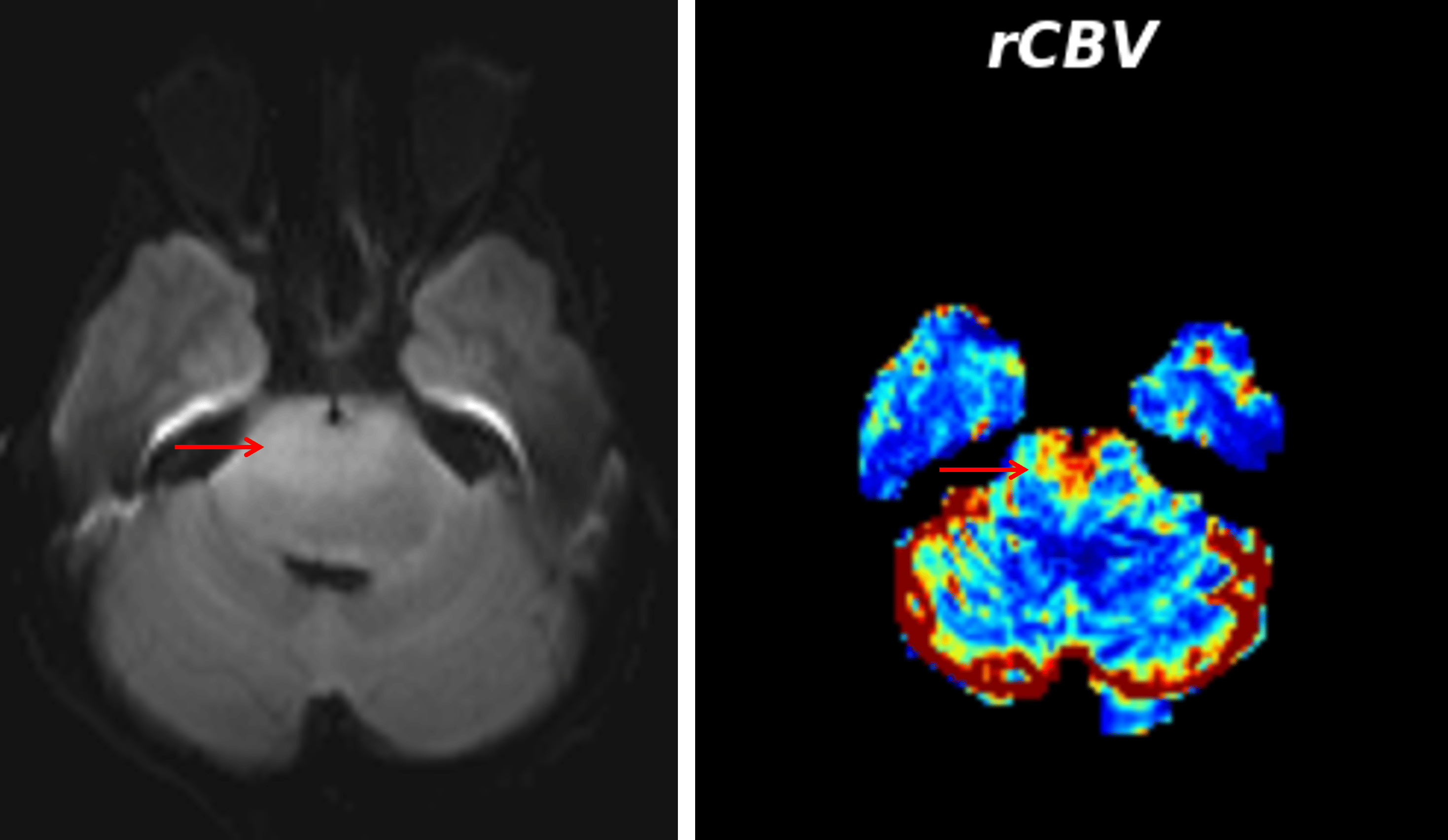
Area of mild restricted diffusion in the ventral pons, which demonstrated elevated cerebral blood volume on perfusion analysis (red arrows).
Diagnosis
Diffuse midline glioma
Key Imaging Features
Become a PRO member to unlock the key imaging features
Differential Diagnosis
Become a PRO member to unlock the differential diagnosis
Discussion
Pearls
Become a PRO member to unlock the pearls