Demographics:
23 years old, Male
Indication:
First time seizure
Findings
CT
- Mixed attenuation mass with internal calcification centered in the posterior right temporal lobe
- Associated mass effect on the right lateral ventricle without midline shift or hydrocephalus
- Scalloping of the adjacent inner table of the calvarium
MRI
- Mixed cystic/solid mass centered in the posterior right temporal lobe measuring 4.5 x 4 x 3.7 cm
- The solid component avidly enhances and the cystic components demonstrate peripheral enhancement
- The more superior of the two dominant cystic components demonstrates relative T1 signal hyperintensity internally
- Areas of susceptibility artifact corresponding with areas of calcification on the CT
- The mass broadly contacts the overlying dura and the ependymal margin of the atrium of the right lateral ventricle, appearing to contact the choroid plexus
- Mild surrounding vasogenic edema
Annotated Images & Illustrations
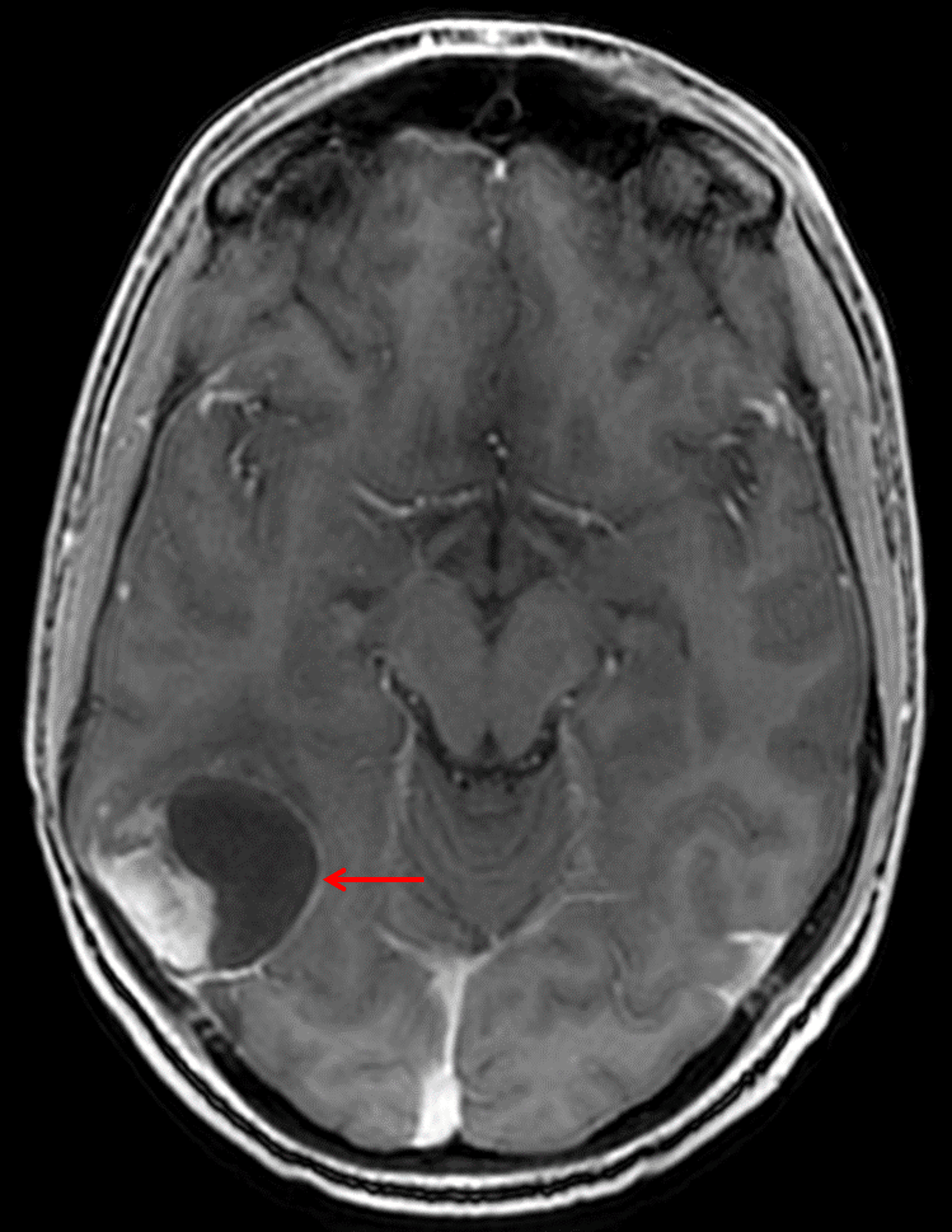
Mixed solid and cystic mass centered in the posterior right temporal lobe (red arrow) with adjacent broad dural contact.
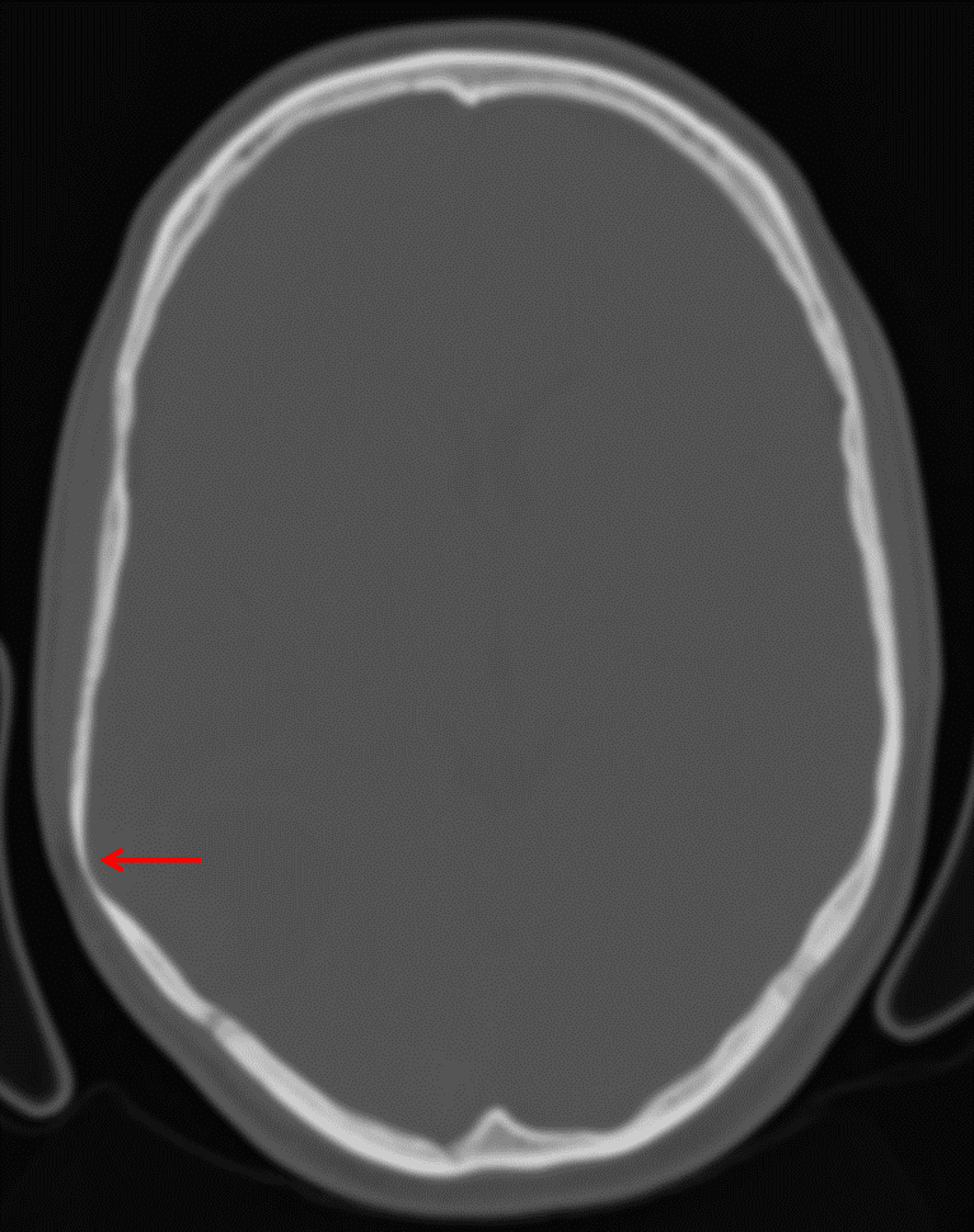
Note overlying smooth scalloping of the inner table of the calvarium (red arrow), favoring this to represent a slow-growing tumor.
Diagnosis
Pleomorphic xanthoastrocytoma (PXA)
Key Imaging Features
Become a PRO member to unlock the key imaging features
Differential Diagnosis
Become a PRO member to unlock the differential diagnosis
Discussion
Pearls
Become a PRO member to unlock the pearls