Findings
CT
- Large, mildly hyperattenuating extra-axial mass centered at midline in the anterior cranial fossa
- Corresponding calcification and adjacent hyperostosis along the olfactory groove
- Vasogenic edema in the adjacent left frontal lobe
- Associated mass effect on the frontal lobes, genu of the corpus callosum, and frontal horns of the lateral ventricles without herniation or hydrocephalus
MRI
- T1 hypointense, T2 isointense extra-axial mass centered in the anterior cranial fossa overlying the olfactory groove
- Diffuse enhancement with radiating internal vascularity
- Diffuse internal restricted diffusion
- Areas of susceptibility artifact corresponding with the areas of calcification seen on CT
- T2/FLAIR signal hyperintensity in the adjacent left frontal white matter
- Posterior displacement of the anterior cerebral arteries
- Possible contacting of the prechiasmatic optic nerves along the inferior margin of the mass
Annotated Images & Illustrations
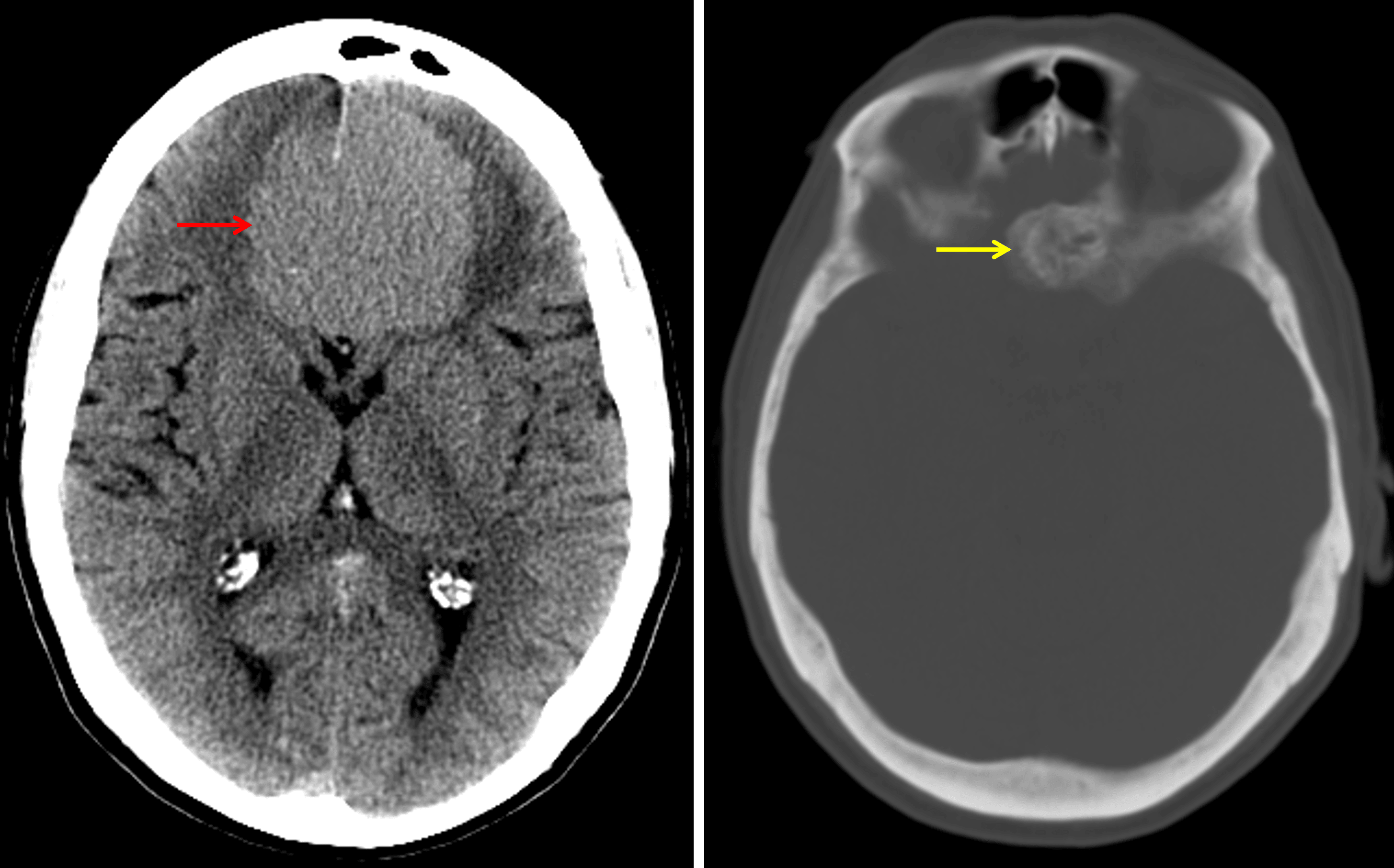
Typical CT appearance of an olfactory groove meningioma, which is hyperattenuating relative to the brain parenchyma (red arrow) and demonstrates adjacent hyperostosis (yellow arrow).
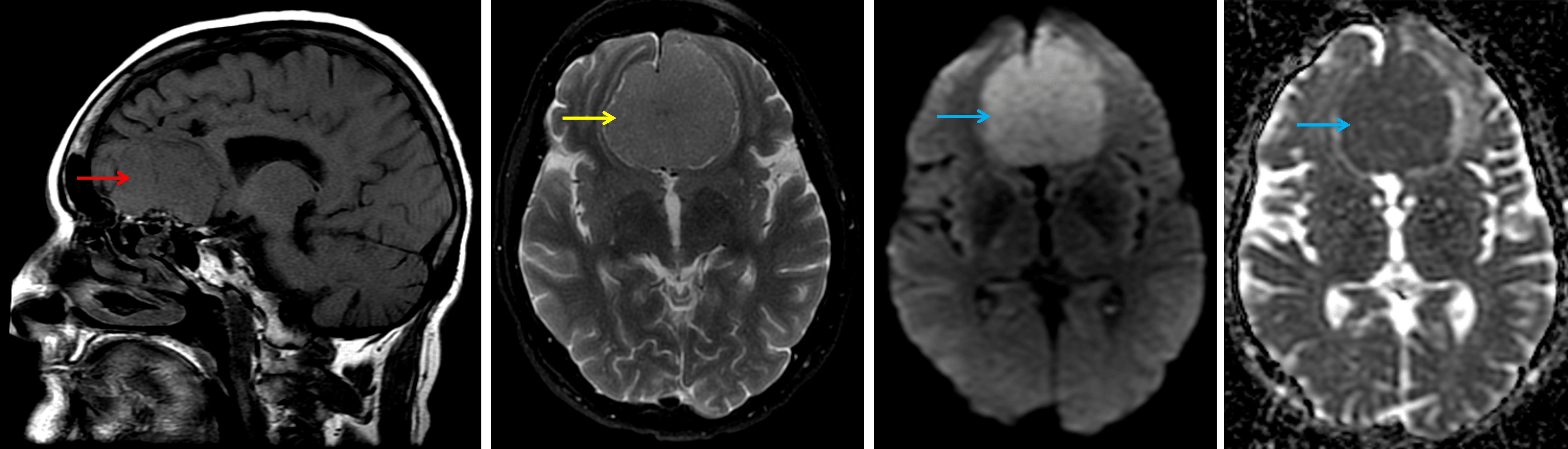
Typical MRI appearance of an olfactory groove meningioma, which is relatively isointense on T1 (red arrow) and T2 (yellow arrow) and demonstrates restricted diffusion (blue arrows).
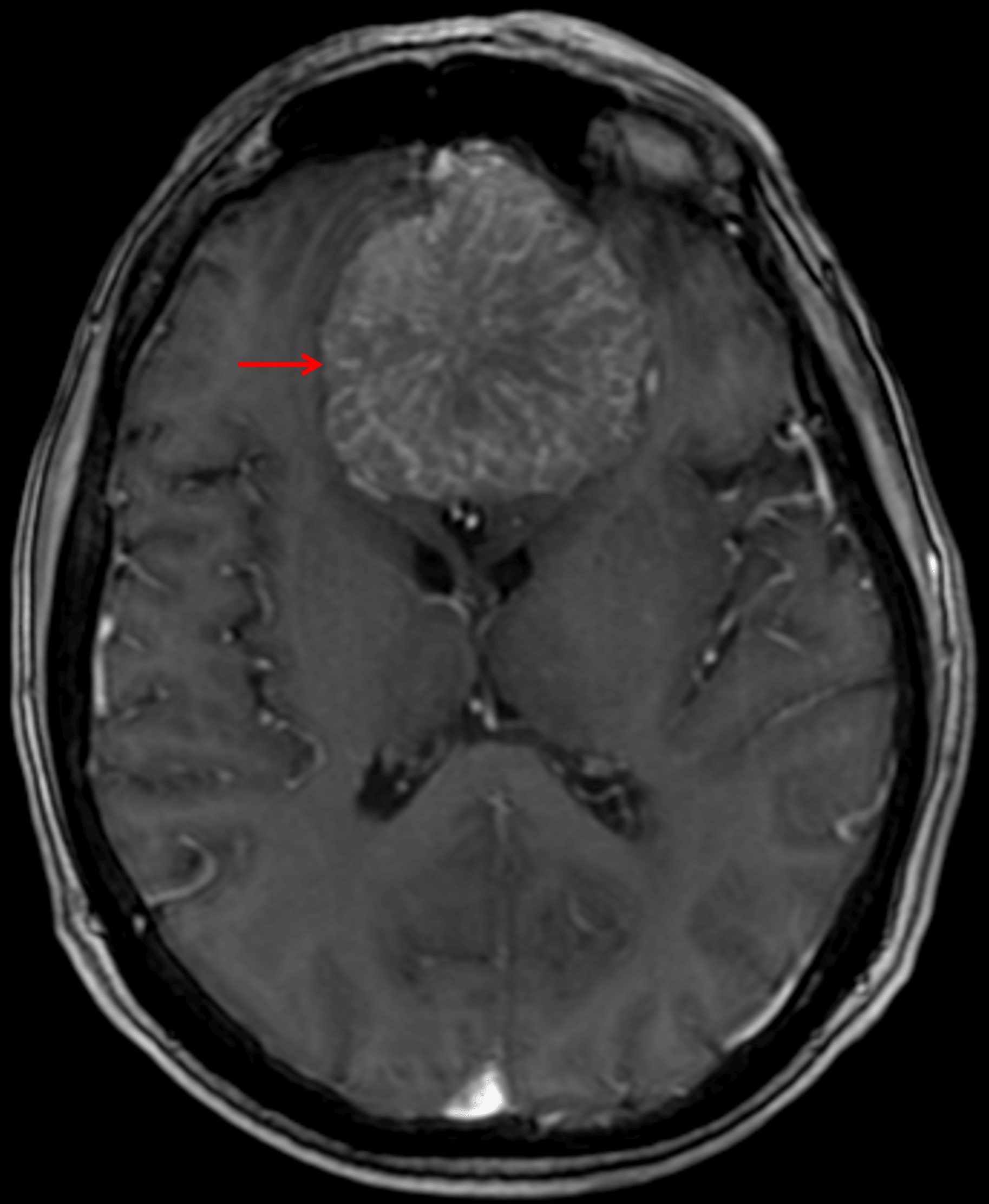
Internal spoke-wheel vascularity on postcontrast imaging (red arrow).
Diagnosis
Meningioma
Key Imaging Features
Become a PRO member to unlock the key imaging features
Differential Diagnosis
Become a PRO member to unlock the differential diagnosis
Discussion
Pearls
Become a PRO member to unlock the pearls
References