Demographics:
51 years old, Male
Indication:
Headache
Findings
CT
- Hyperattenuating extra-axial mass overlying the left sylvian fissure with small internal areas of hypoattenuation and surrounding vasogenic edema
- Lytic changes involving the inner table of the overlying calvarium
MRI
- T1 isointense, mildly T2 hyperintense, diffusely enhancing extra-axial mass overlying the left sylvian fissure measuring 6 x 4.2 x 6.4 cm
- Small internal areas of more pronounced T2 signal hyperintensity
- Mild corresponding restricted diffusion throughout the mass
- Surrounding vasogenic edema
- Associated mass effect resulting in local sulcal effacement, crowding of the left lateral ventricle, 7 mm left-to-right midline shift, and medialization of the left temporal uncus with crowding of the basal cisterns and mass effect on the midbrain
Annotated Images & Illustrations
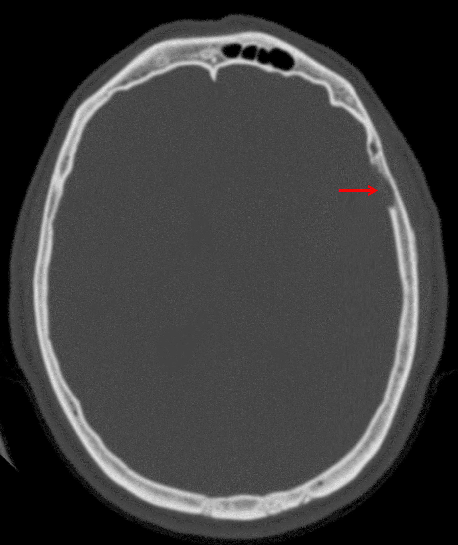
Inner table calvarial erosion overlying the mass (red arrow).
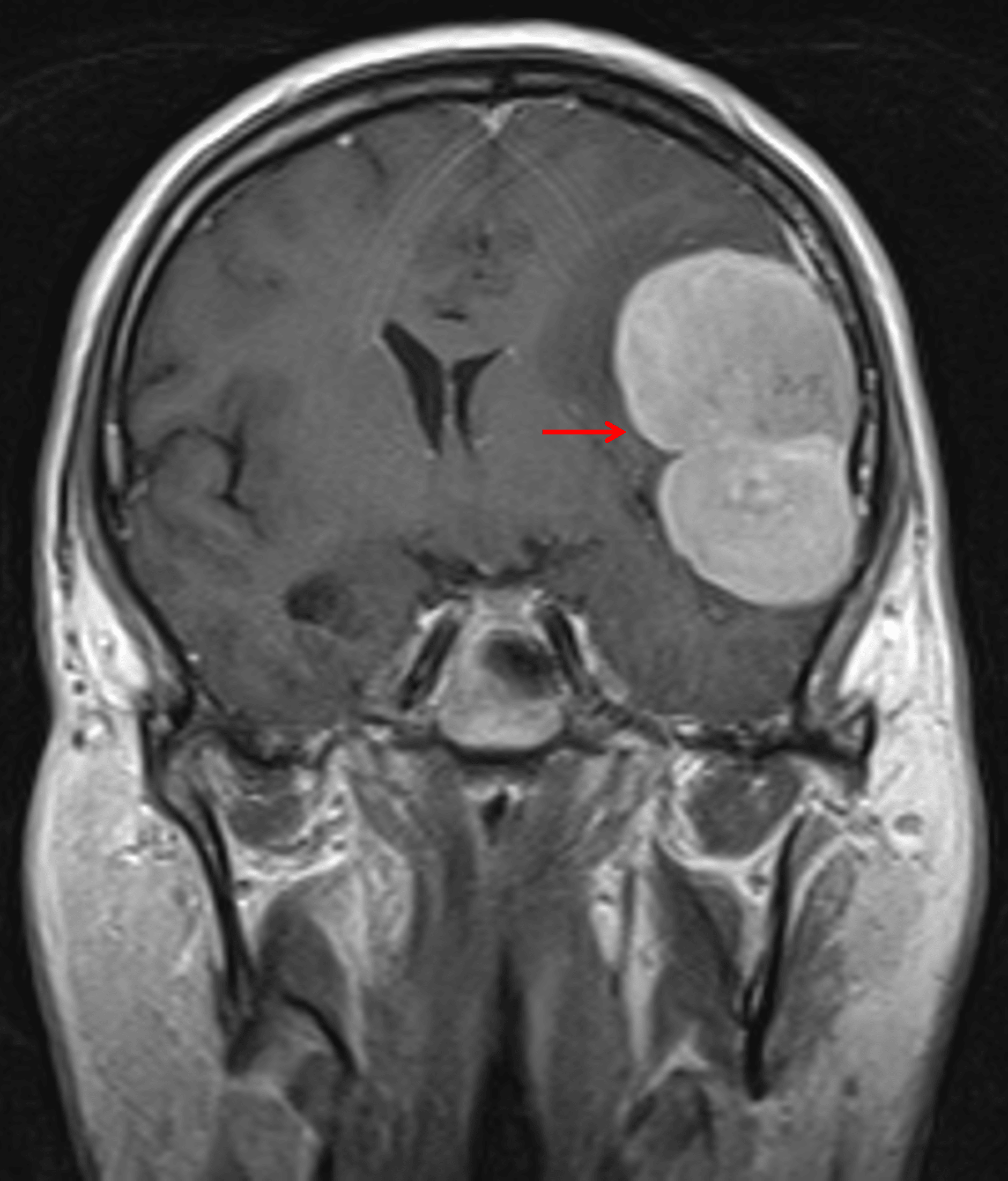
Avidly enhancing mass with lobulated contours (red arrow).
Diagnosis
Solitary fibrous tumor (hemangiopericytoma)
Key Imaging Features
Become a PRO member to unlock the key imaging features
Differential Diagnosis
Become a PRO member to unlock the differential diagnosis
Discussion
Pearls
Become a PRO member to unlock the pearls
References