Findings
- Heterogeneous mass in the left cerebral hemisphere measuring 5.3 x 5 x 6 cm
- Corresponding heterogeneous enhancement with matching areas of restricted diffusion
- Areas of intrinsic T1 signal hyperintensity, particularly along the deep margin of the mass
- Surrounding vasogenic edema and associated mass effect resulting in local sulcal effacement, crowding of the left lateral ventricle, and 10 mm left-to-right midline shift
- Entrapment of the temporal horn of the left lateral ventricle
Annotated Images & Illustrations
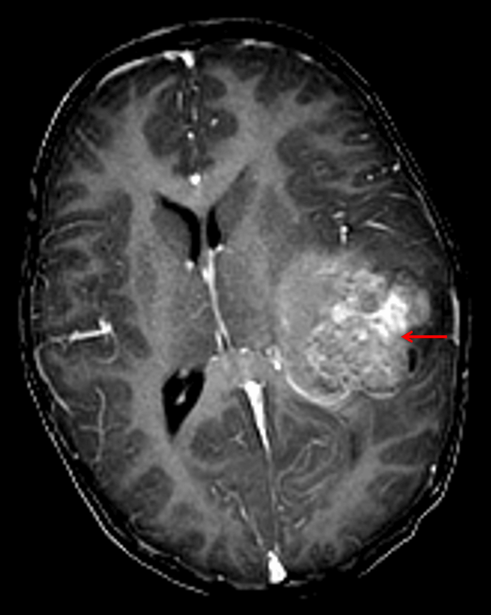
Heterogeneously enhancing mass in the left cerebral hemisphere (red arrow) with associated local mass effect and midline shift.
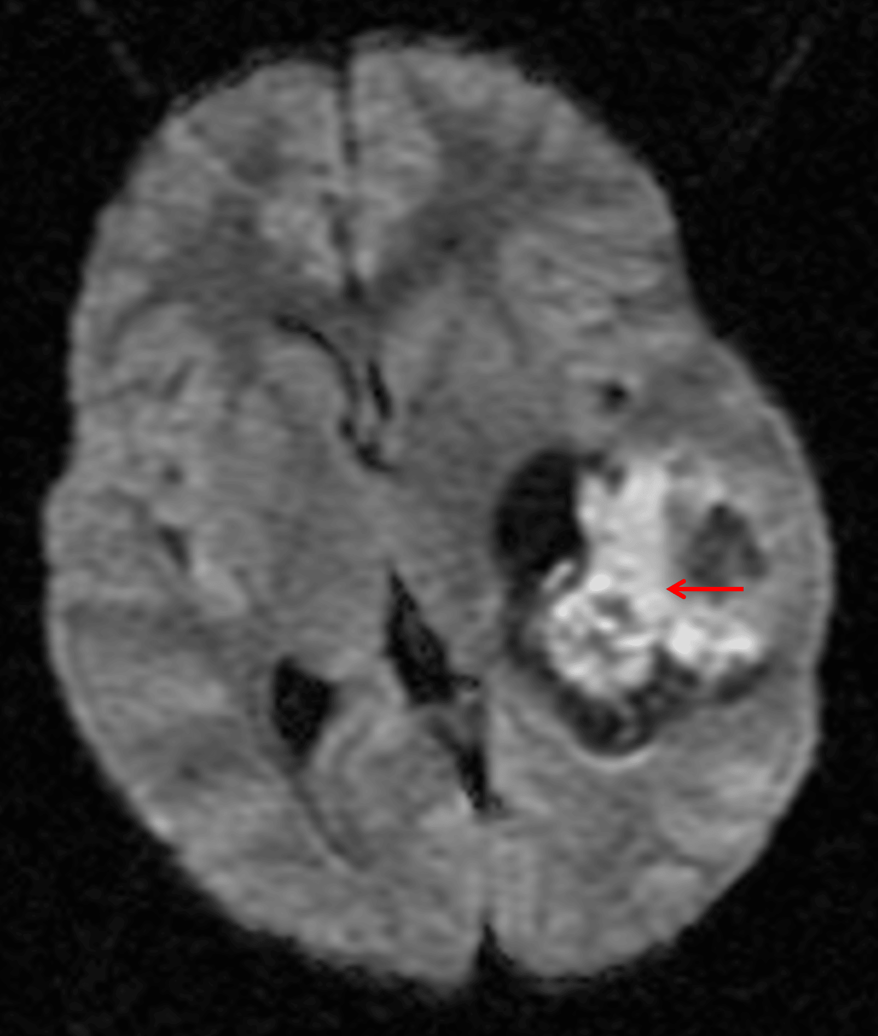
Areas of internal restricted diffusion (red arrow) corresponding with the enhancing components, indicative of hypercellularity.
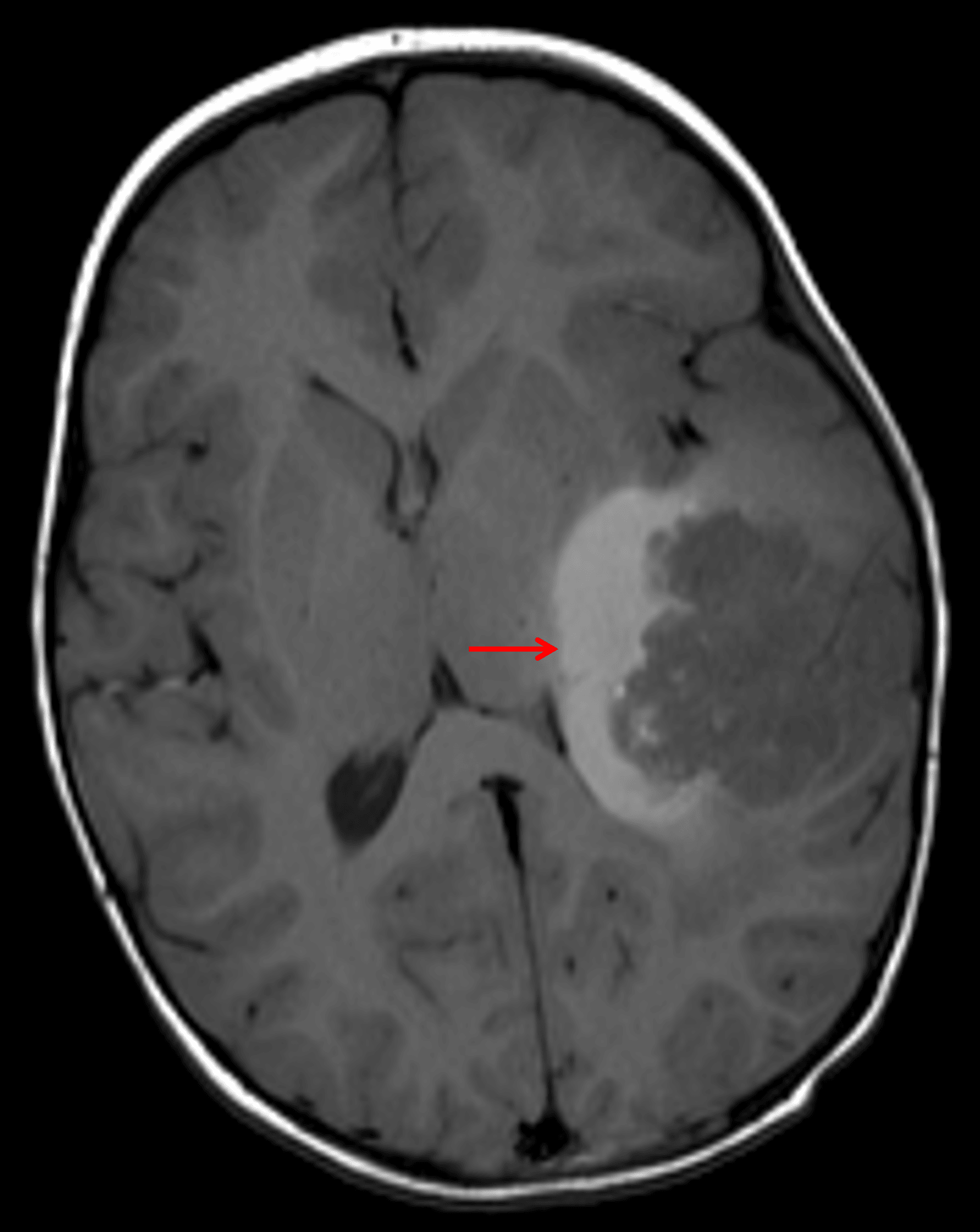
Area of T1 signal hyperintensity along the deep margin of the tumor (red arrow), likely representing tumor-related hemorrhage.
Diagnosis
Atypical teratoid/rhabdoid tumor (AT/RT)
Key Imaging Features
Become a PRO member to unlock the key imaging features
Differential Diagnosis
Become a PRO member to unlock the differential diagnosis
Discussion
Pearls
Become a PRO member to unlock the pearls