Findings
- Large, heterogeneous mass centered in the quadrigeminal plate region measuring 6 x 2.8 x 6 cm
- Extensive corresponding mass effect on the midbrain with effacement of the cerebral aqueduct and associated severe obstructive hydrocephalus with cerebral parenchymal thinning
- Small volume hemorrhage layering in both lateral ventricles and fourth ventricle with rounded clot in the third ventricle
- Small volume scattered subarachnoid hemorrhage
- Additional noncontiguous masses in the left cerebellopontine angle cistern and along the ependymal margin of the frontal horn of the left lateral ventricle at the caudothalamic groove
- The above described masses demonstrate areas of restricted diffusion (particularly the two smaller lesions) but no discrete enhancement
Annotated Images & Illustrations
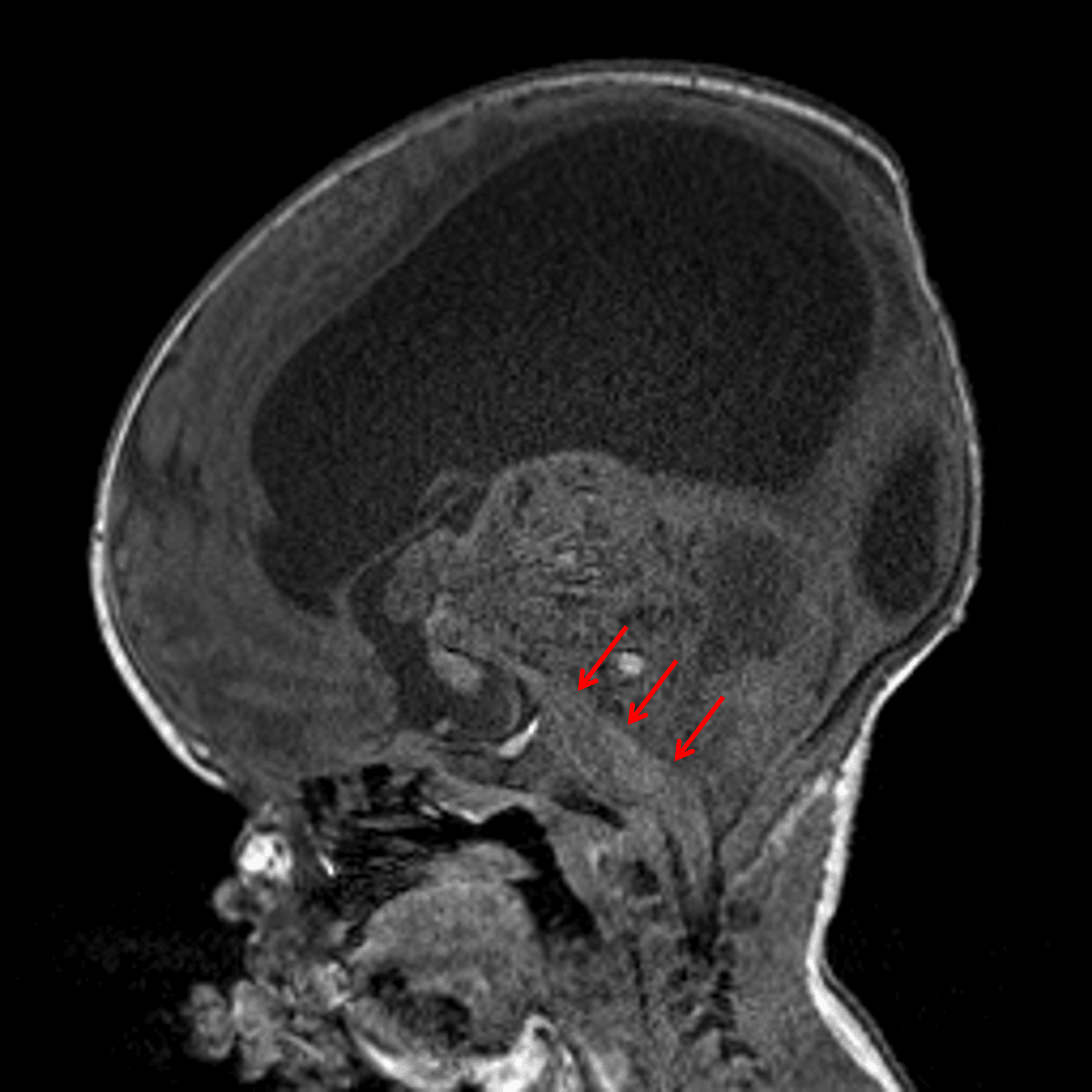
Large mass centered in the superior aspect of the posterior fossa with anterior displacement of and severe mass effect on the brainstem (red arrows).
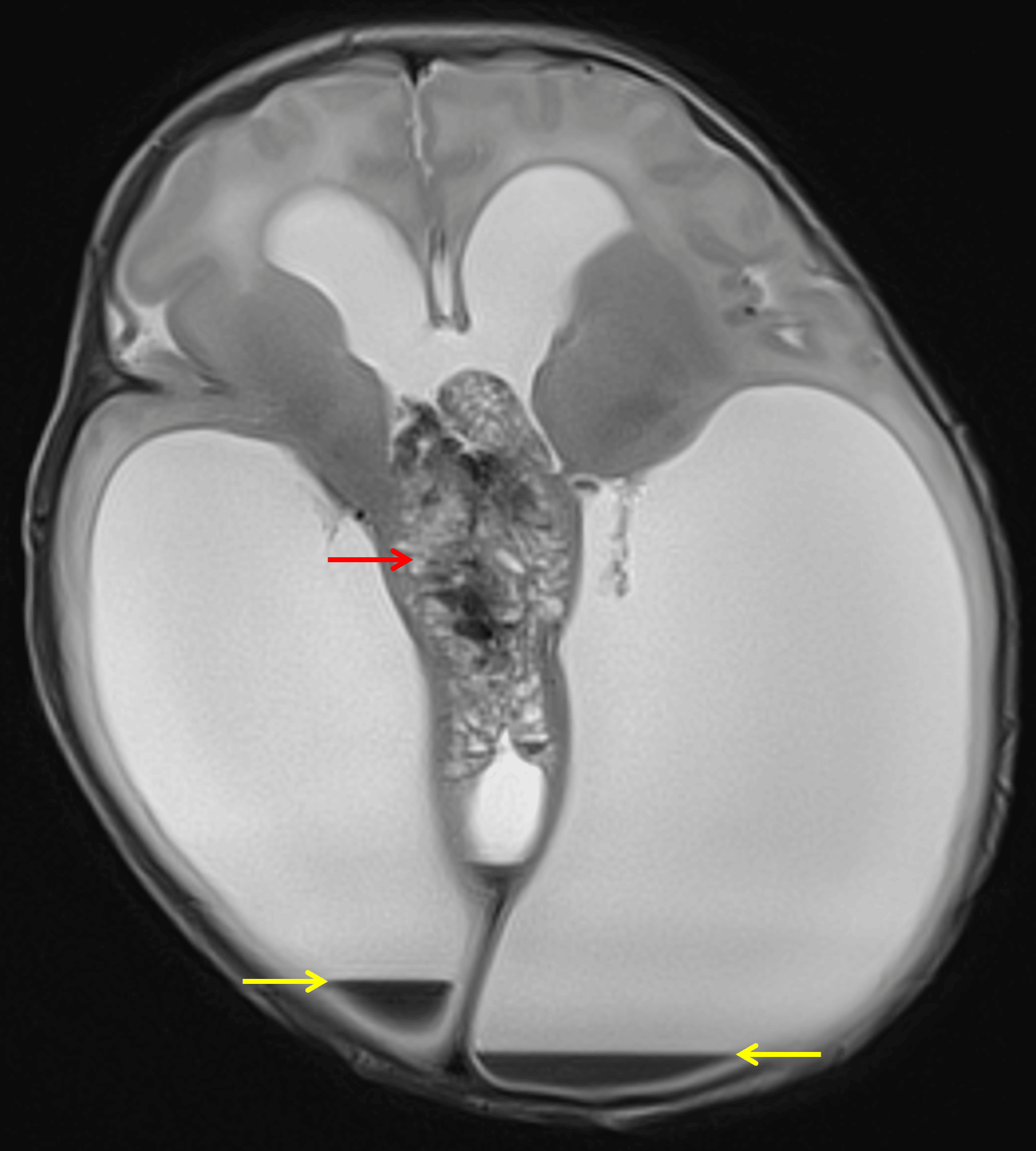
Heterogeneous midline mass (red arrow) with associated severe obstructive hydrocephalus and small volume intraventricular hemorrhage (yellow arrows).
This mass demonstrates no appreciable corresponding enhancement.
Diagnosis
Embryonal tumor with multilayered rosettes (ETMR)
Key Imaging Features
Become a PRO member to unlock the key imaging features
Differential Diagnosis
Become a PRO member to unlock the differential diagnosis
Discussion
Pearls
Become a PRO member to unlock the pearls