Use mouse wheel, arrow keys or left click (with scroll tool selected) to scroll
ui.case.use_touch_gestures
DICOM HelpSource: Local (us-east1-c)
Keyboard shortcuts (Alt+K)
Demographics:
33 years old, Female
Indication:
Shortness of breath
Findings
- Blurring of the right heart border on the PA projection with a triangular opacity seen overlying the cardiac silhouette on the lateral projection
- Right IJ approach port catheter with tip projecting over the superior cavoatrial junction
- No pleural effusion or pneumothorax
- Normal size and configuration of the cardiopericardial silhouette
- Emphysema
Diagnosis
Right middle lobe collapse
Sample Report
Right middle lobe collapse. Recommend followup imaging to ensure resolution. If there is persistent lobar collapse on followup imaging, chest CT should be obtained to assess for a fixed obstructive lesion.
Discussion
- Right middle lobe collapse classically results in a triangular anterior opacity on the lateral view and hazy opacity with loss of the right heart border on frontal view
- Look for evidence of volume loss when trying to decide between atelectasis and consolidation. Signs of volume loss include hemidiaphragm elevation and hilar displacement toward the abnormality
Annotated Images & Illustrations
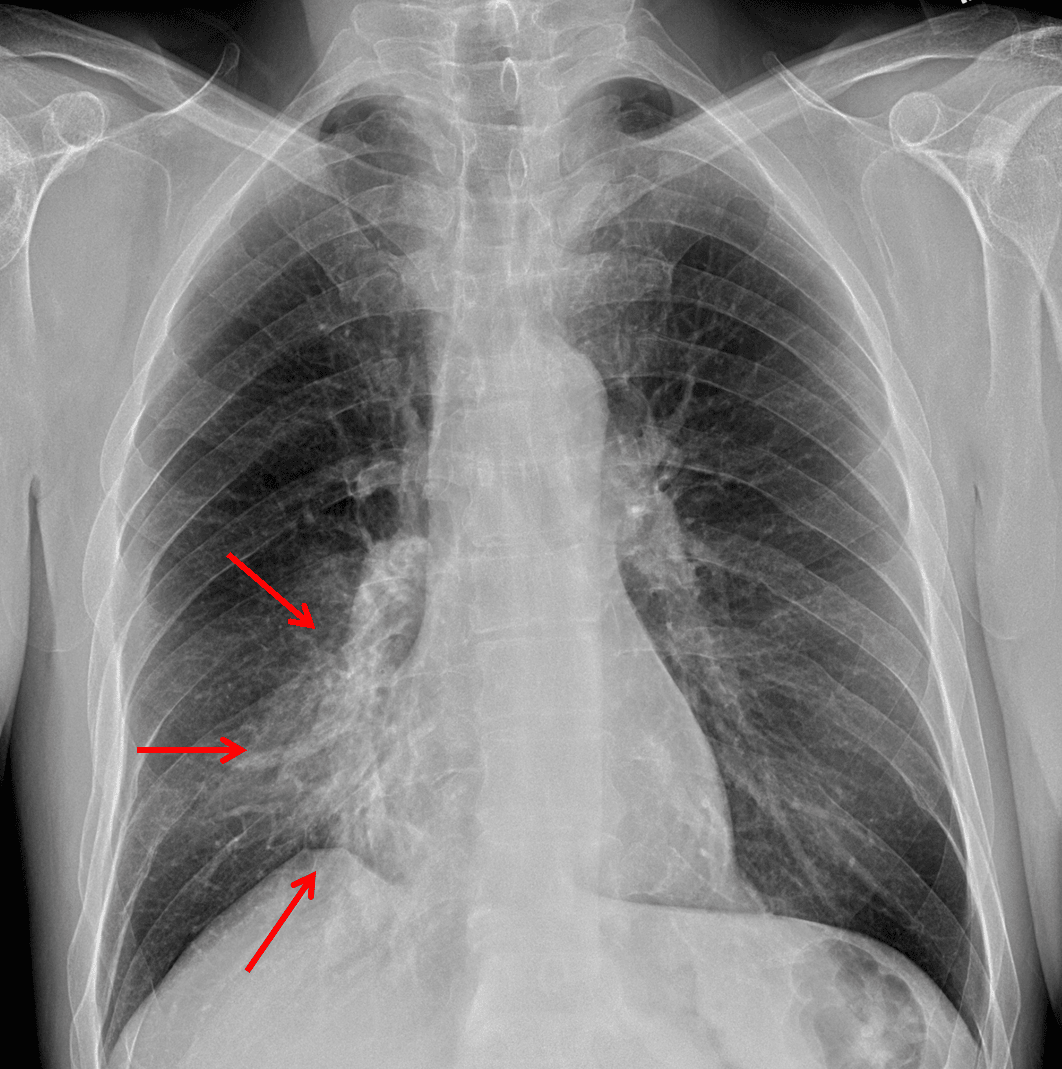
Red arrows: hazy airspace opacity with obscuring of the right heart border.
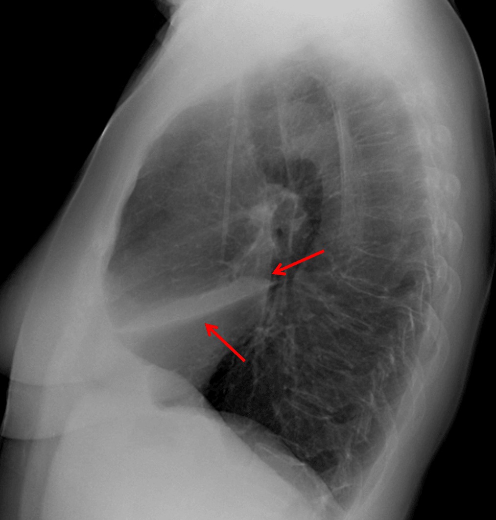
Red arrows: anterior airspace opacity sharply marginated by the major fissure inferiorly.
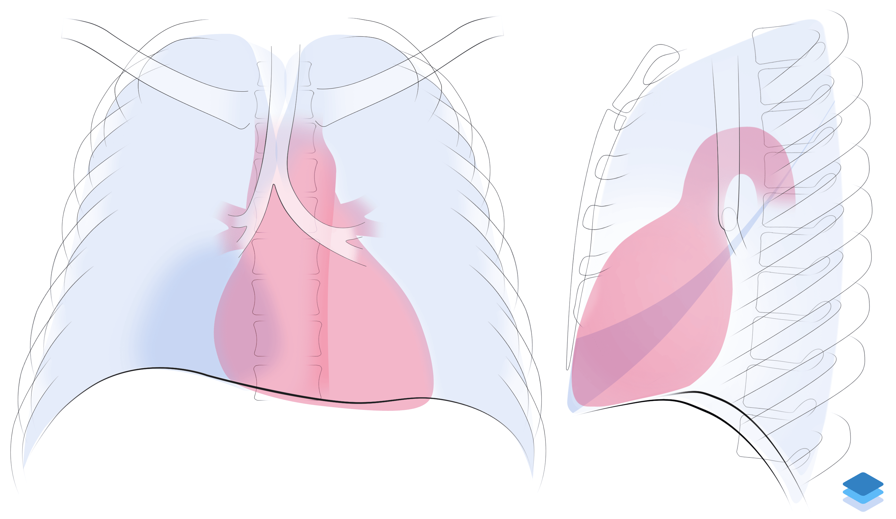
Right middle lobe collapse. Illustration by Valerie George, MD