患者信息:
33 岁, 女性
检查指征:
呼吸困难
诊断
右中叶肺不张
示例报告
右中叶肺不张。建议随访影像检查以确认消散。如随访影像显示持续性肺叶不张,应行胸部CT评估是否存在固定性梗阻性病变。
讨论
- 右中叶不张的经典表现是侧位片上前方三角形密度影和正位片上右心缘模糊的雾状影
- 在鉴别不张和实变时注意观察容积减小的证据。容积减小的征象包括同侧膈肌抬高和肺门向病变方向移位
带注释的图像和插图
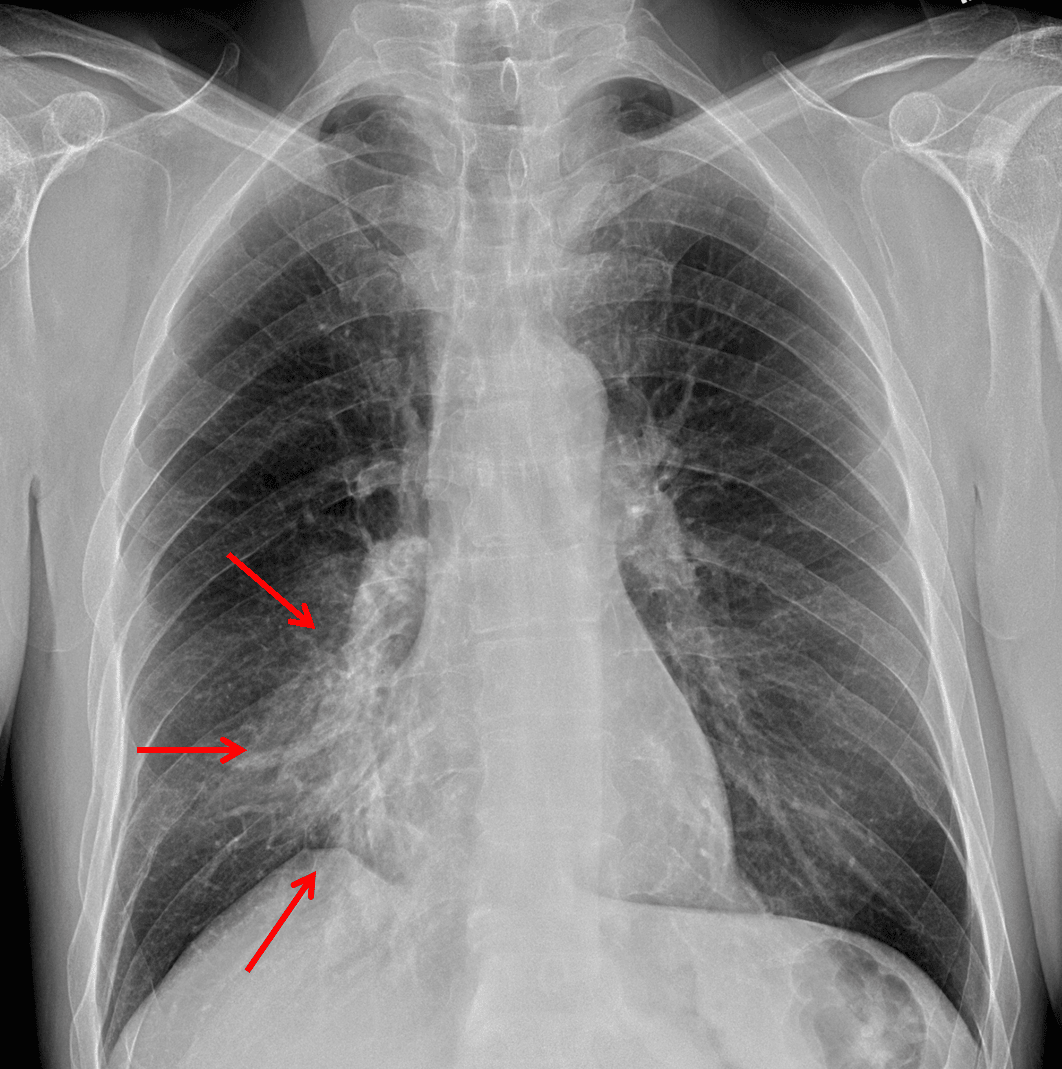
Red arrows: hazy airspace opacity with obscuring of the right heart border.
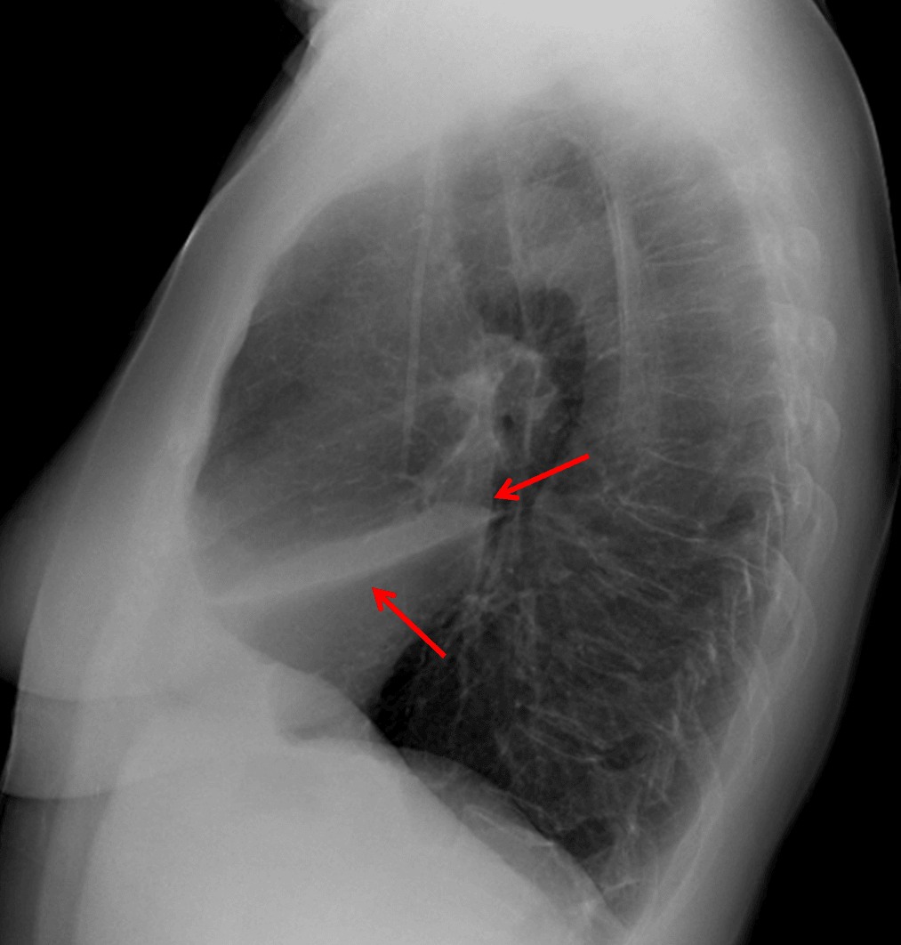
Red arrows: anterior airspace opacity sharply marginated by the major fissure inferiorly.
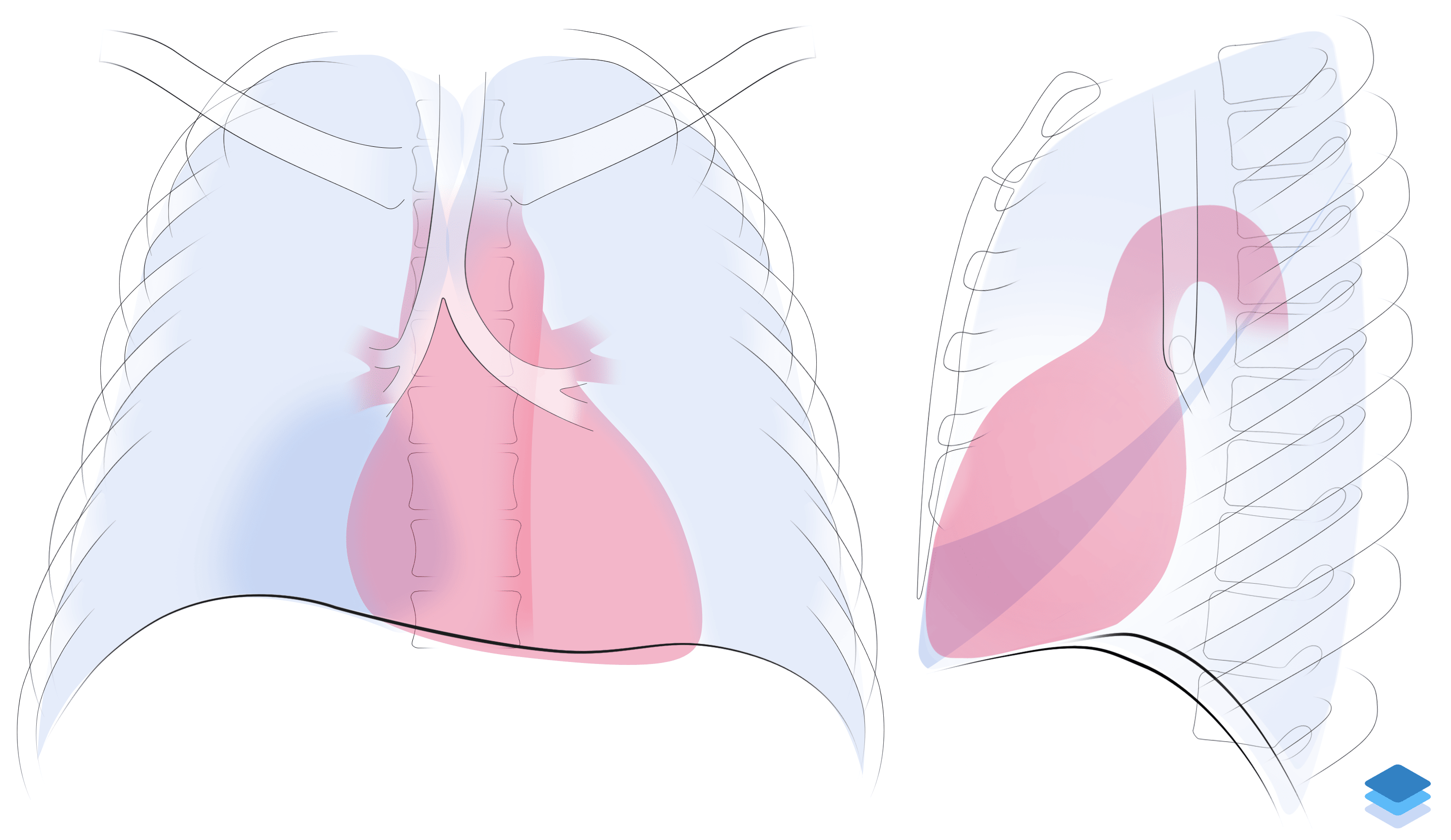
Right middle lobe collapse. Illustration by Valerie George, MD