病例 #1
影像所见
- 右肺上叶密度增高及容积缩小,副裂向上移位
- 纵隔轻度右移
- 左肺下叶轻度斑片状密度增高影
- 右侧颈内静脉入路Port-A-Cath,尖端位于上腔静脉-右心房交界处
- 无胸腔积液或气胸
- 心包影大小及形态正常
诊断
右肺上叶不张
示例报告
右肺上叶不张,需警惕黏液栓塞或中央阻塞性肿块。建议行胸部CT进一步评估。
左肺下叶轻度斑片状影为非特异性表现,可能与肺不张、误吸或肺炎相关。
讨论
- 右肺上叶不张通常在正位片上表现为容积缩小和密度增高,伴副裂上移
- Golden S征是由于副裂内侧部分被肺门肿块牵拉固定,而中间及外侧部分上移所致——因此如果看到这种表现,应考虑中央阻塞性肿块
带注释的图像和插图
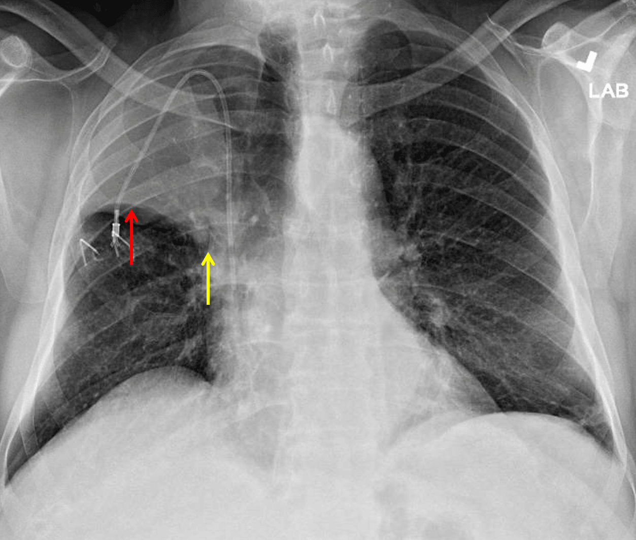
Right upper lobe collapse. The minor fissure is elevated laterally (red arrow) but not as elevated centrally (yellow arrow) raising concern for a perihilar mass.
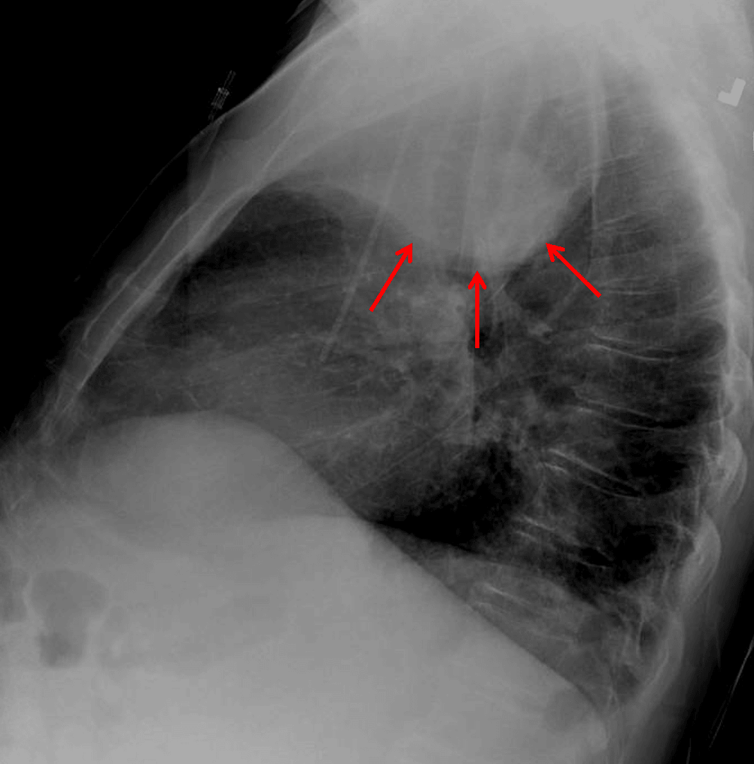
Right upper lobe collapse. Lateral view shows a paradoxical bulging appearance along the inferior margin of the collapsed lobe concerning for mass (red arrows).
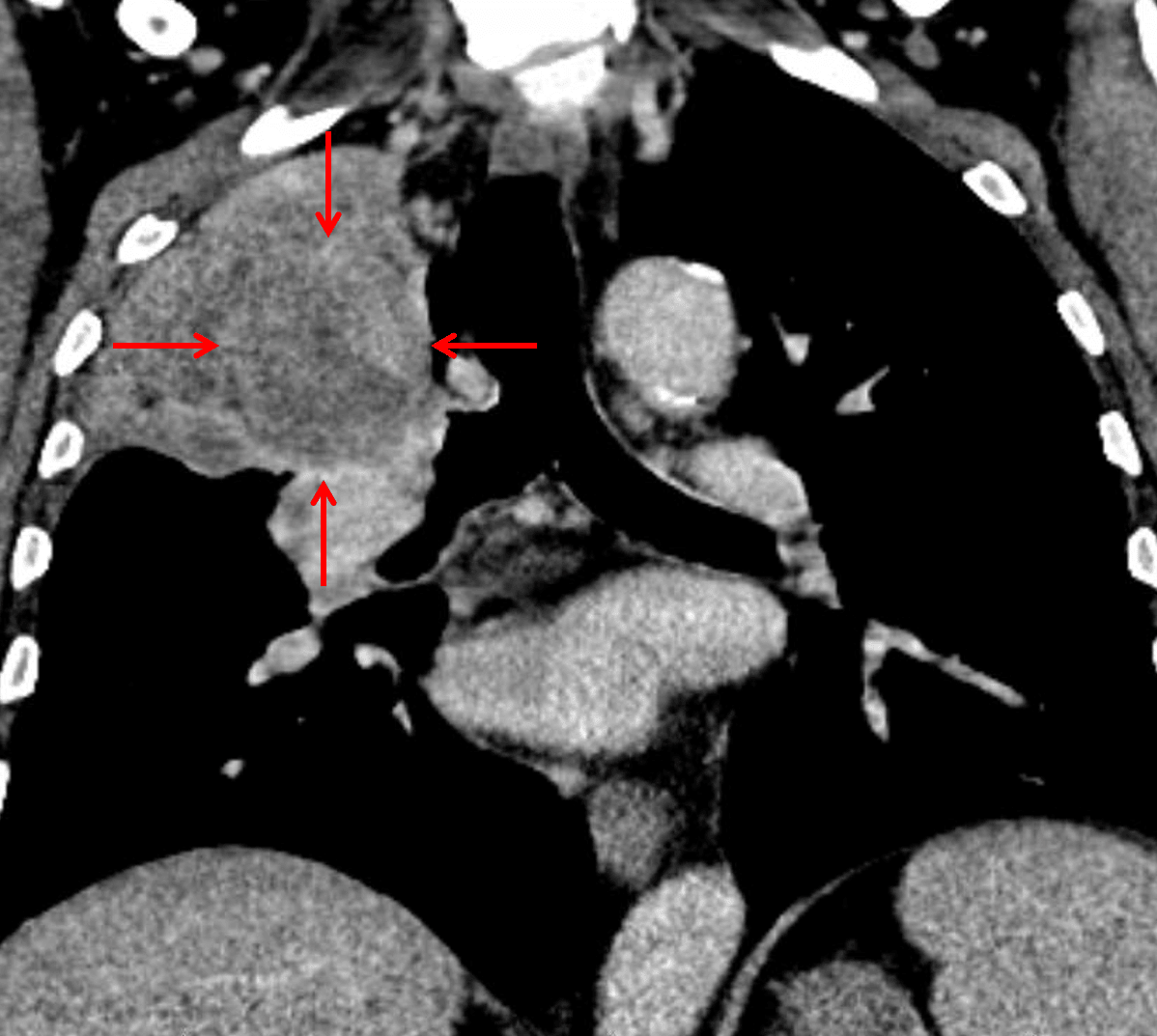
Subsequent CT confirmed the presence of a suprahilar mass with bronchial invasion (red arrows), though the mass is difficult to distinguish from the adjacent atelectatic lung.
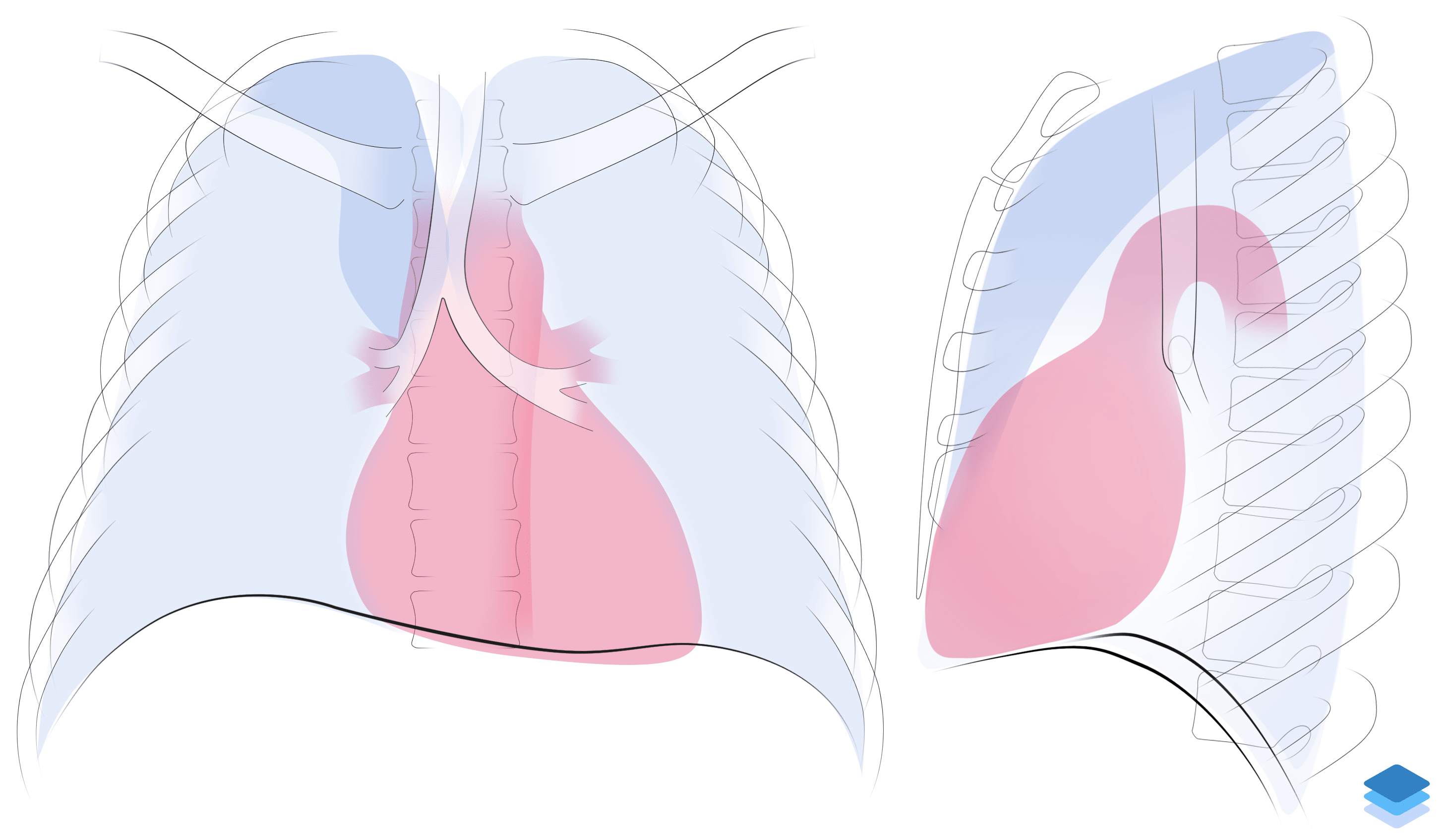
Right upper lobe collapse. Illustration by Valerie George, MD