Demographics:
68 years old, Female
Indication:
Aphasia
Findings
CT
- Area of hypoattenuation in the left frontal subcortical and deep white matter
MRI
- Relatively rounded area of T2/FLAIR signal hyperintensity in the left frontal white matter
- Corresponding irregular, discontinuous areas of curvilinear enhancement along the periphery of the lesion
- Diffuse corresponding restricted diffusion with the exception of a small rounded area of facilitated diffusion centrally
- No corresponding susceptibility artifact
- No substantial corresponding mass effect
Annotated Images & Illustrations
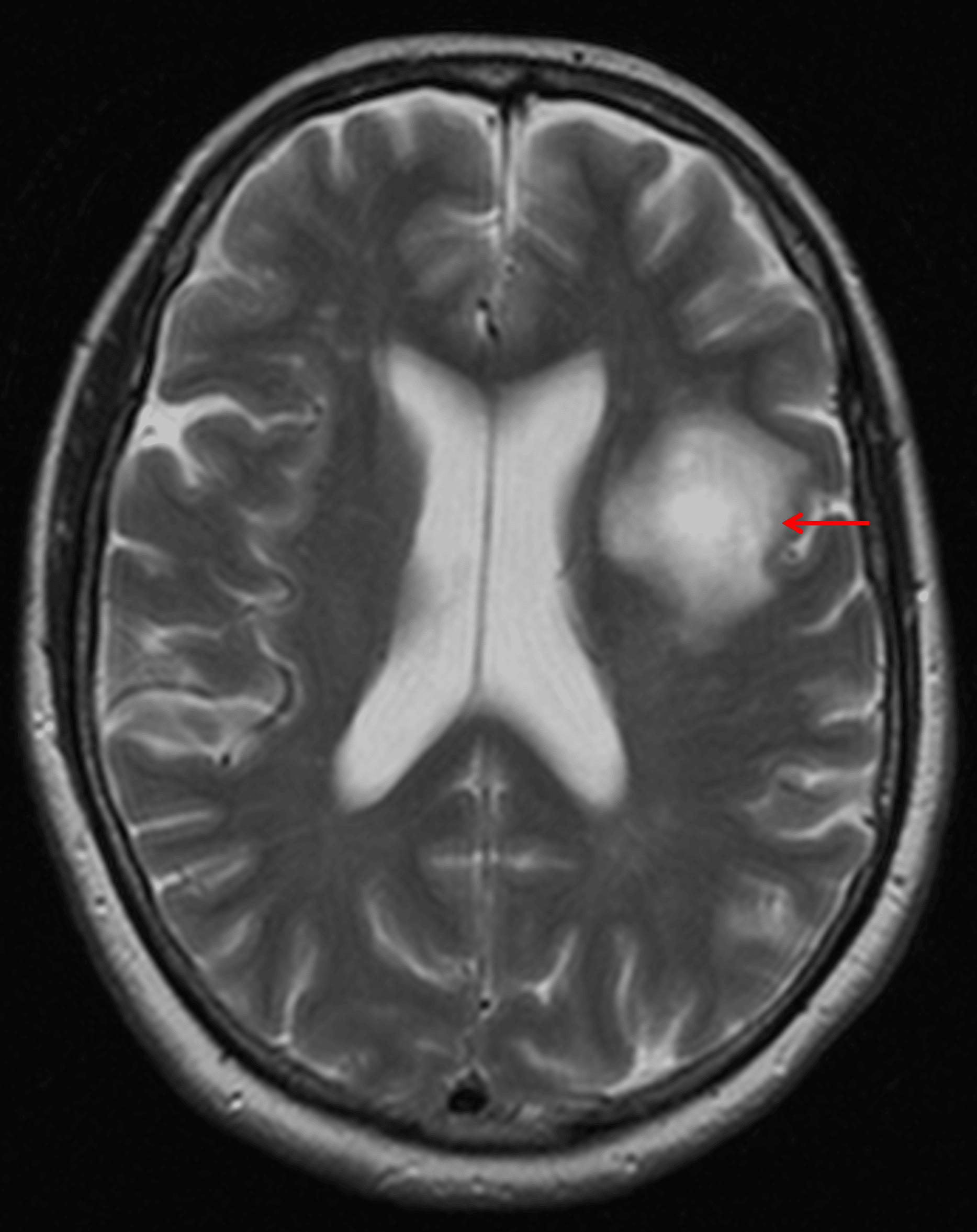
Masslike area of T2 signal hyperintensity in the left frontal white matter (red arrow) with surprisingly essentially no mass effect on the left lateral ventricle or adjacent sulci.
Diagnosis
Tumefactive demyelinating lesion
Key Imaging Features
Become a PRO member to unlock the key imaging features
Differential Diagnosis
Become a PRO member to unlock the differential diagnosis
Discussion
Pearls
Become a PRO member to unlock the pearls