Demographics:
3 years old, Female
Indication:
Gait instability, confusion
Findings
CT
- Mildly hyperattenuating mass in the left cerebellar hemisphere with mass effect on the fourth ventricle and resultant obstructive hydrocephalus
- No corresponding calcification
MRI
- T1 hypointense, slightly T2 hyperintense mass centered in the inferior aspect of the left cerebellar hemisphere with diffuse enhancement and mild diffuse restricted diffusion
- Crescentic cystic components along the periphery of the mass
- Mild adjacent parenchymal edema
- Corresponding mass effect with obstructive hydrocephalus at the level of the fourth ventricle and mild upward transtentorial herniation
Annotated Images & Illustrations
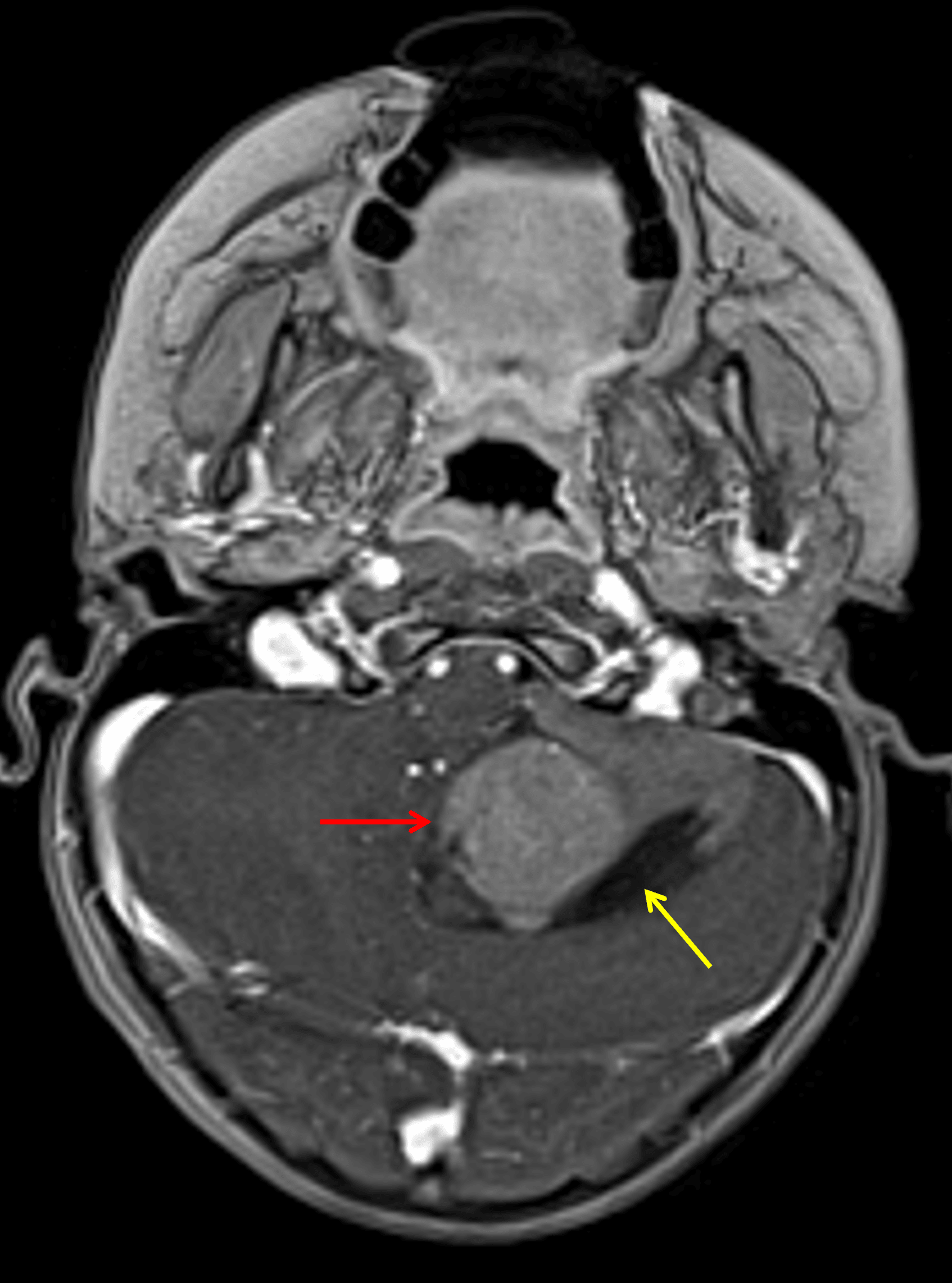
Enhancing mass centered in the left cerebellar hemisphere (red arrow) with a peripheral cystic component (yellow arrow).
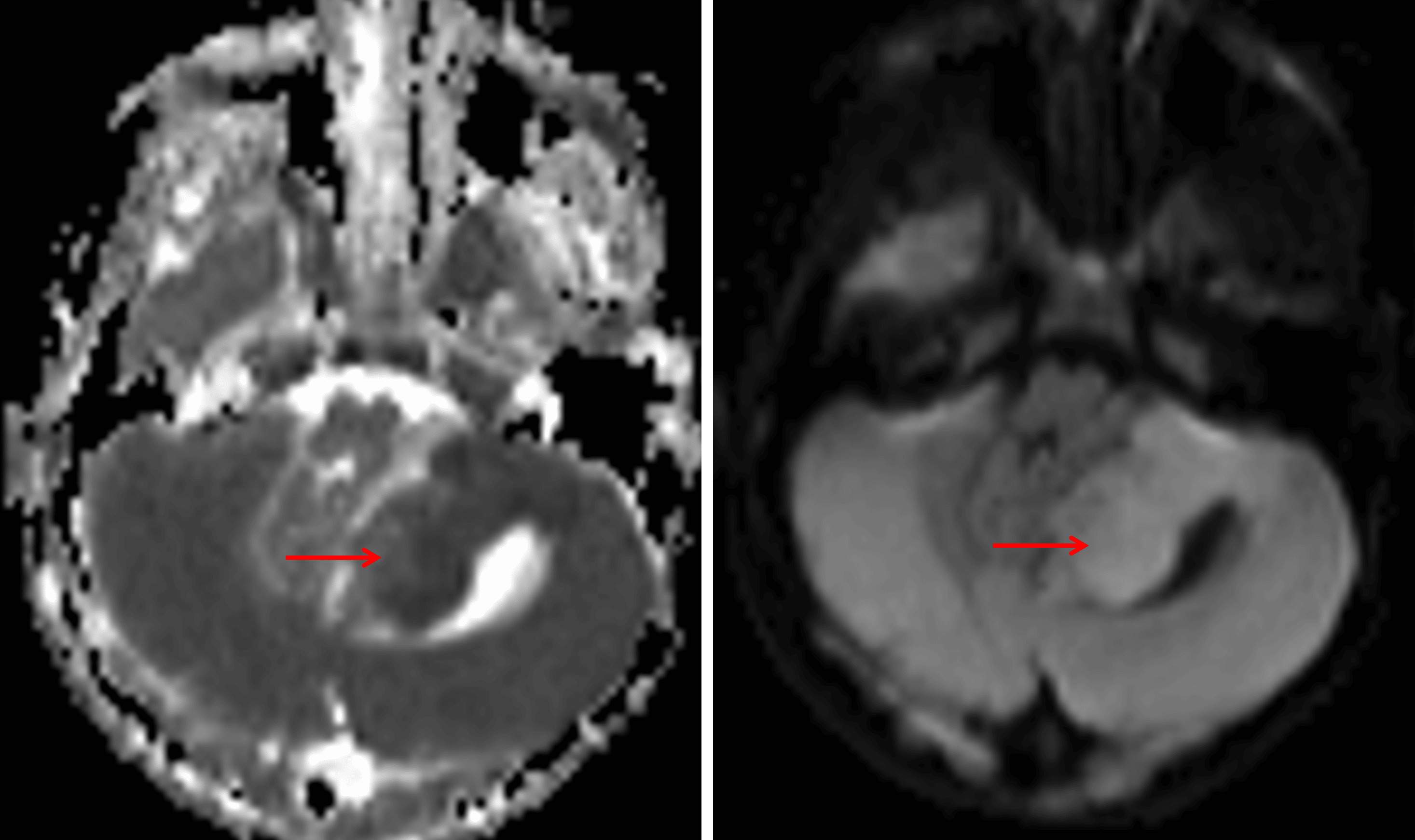
Mild corresponding restricted diffusion (red arrows).
Diagnosis
Medulloblastoma (SHH-activated, TP53 wild-type, nodular desmoplastic histology)
Differential Diagnosis
Become a PRO member to unlock the differential diagnosis
Pearls
Become a PRO member to unlock the pearls