Demographics:
17 years old, Female
Indication:
Encephalopathy
Findings
- T2/FLAIR signal hyperintensity in the right greater than left thalami and right greater than left dorsal midbrain with involvement of the periaqueductal gray matter, hypothalamus, anterior commissure, and mamillary bodies
- Areas of cystic change in the right thalamus and right eccentric midbrain
- No corresponding enhancement or restricted diffusion
- No substantial intracranial mass effect or evidence of hydrocephalus
Annotated Images & Illustrations
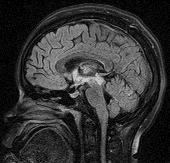
FLAIR signal hyperintensity along the ependymal margins of the third ventricle and cerebral aqueduct, extending into the right greater than left thalami.
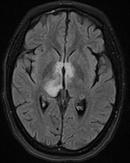
FLAIR signal hyperintensity along the ependymal margins of the third ventricle and cerebral aqueduct, extending into the right greater than left thalami.
Diagnosis
Neuromyelitis optica (NMO)
Key Imaging Features
Become a PRO member to unlock the key imaging features
Differential Diagnosis
Become a PRO member to unlock the differential diagnosis
Discussion
Pearls
Become a PRO member to unlock the pearls