Demographics:
23 years old, Female
Indication:
Double vision
Findings
Initial MRI
- Relatively symmetric, minimally expansile T2/FLAIR signal hyperintensity in the midbrain and pons (predominantly involving the corticospinal and central tegmental tracts with sparing of the transverse pontine fibers) extending superiorly into the right greater than left internal capsules. There is also involvement of the left superior cerebellar peduncle
- No corresponding enhancement, restricted diffusion, or susceptibility artifact
6 month follow-up MRI
- Overall improved appearance and extent of T2/FLAIR signal hyperintensity in the brainstem extending into the internal capsules
- New expansile T2 signal hyperintensity in the right ventral medulla
- No corresponding enhancement, restricted diffusion, or susceptibility artifact
Annotated Images & Illustrations
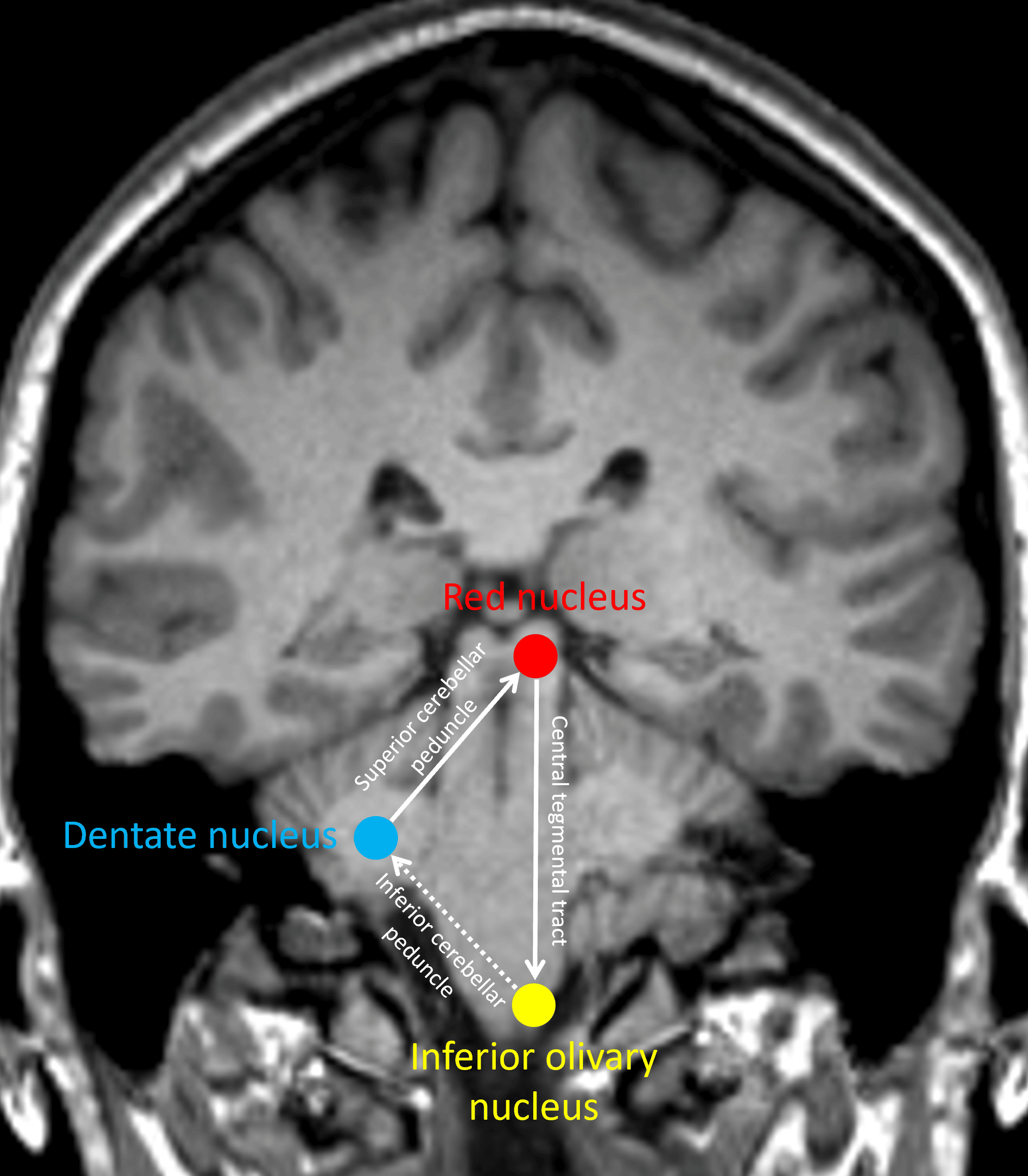
The infamous Guillain-Mollaret triangle.
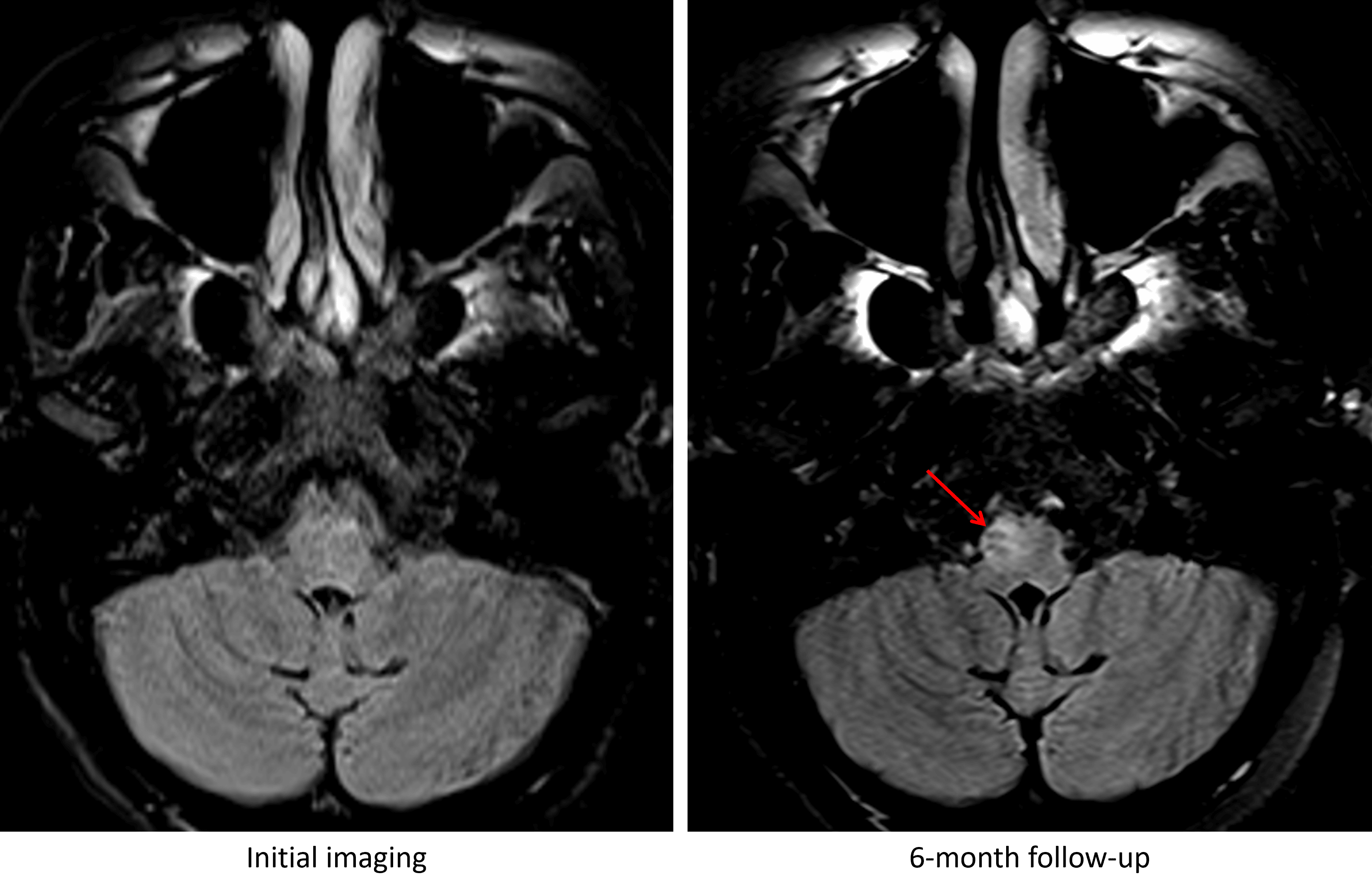
Development of T2/FLAIR signal hyperintensity and enlargement of the right inferior olivary nucleus at 6 month followup imaging (red arrow).
Diagnosis
Hypertrophic olivary degeneration (HOD)
Key Imaging Features
Become a PRO member to unlock the key imaging features
Differential Diagnosis
Become a PRO member to unlock the differential diagnosis
Discussion
Pearls
Become a PRO member to unlock the pearls