Demographics:
52 years old, Male
Indication:
Vision loss
Findings
- Peripherally-enhancing sellar/suprasellar mass measuring 2.5 x 1.6 x 1.7 cm, which is not separable from the pituitary gland
- Centrally, the lesion is mildly T1 hypointense and T2 hyperintense
- No corresponding restricted diffusion
- The lesion bulges to the intercarotid line on the right and remains medial to the medial carotid tangent line on the left
- The lesion contacts and mildly uplifts the optic chiasm
Annotated Images & Illustrations
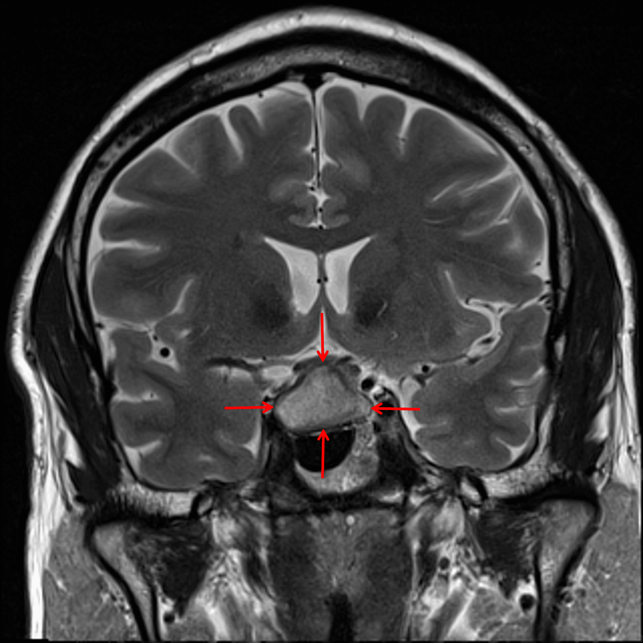
Cystic sellar/suprasellar mass with internal T2 signal hyperintensity (red arrows).
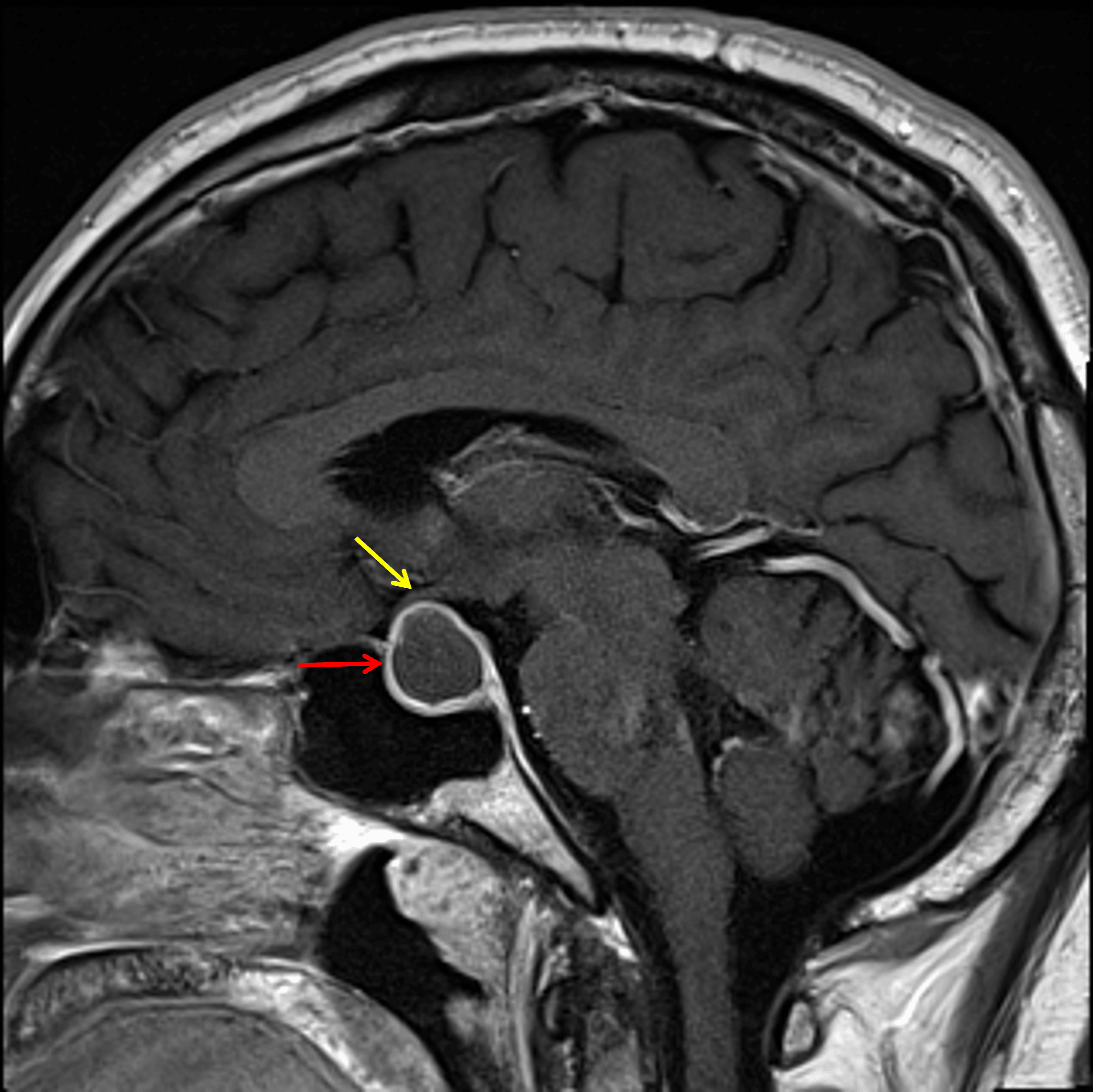
The lesion demonstrates peripheral enhancement (red arrow) and contacts and mildly uplifts the optic chiasm (yellow arrow).
Diagnosis
Cystic pituitary adenoma
Differential Diagnosis
Become a PRO member to unlock the differential diagnosis
Pearls
Become a PRO member to unlock the pearls