Demographics:
43 years old, Male
Indication:
Headache, history of HIV/AIDS
Findings
CT
- Ill-defined hypoattenuating areas involving the left basal ganglia, right thalamus, and bilateral internal capsule
- Internal foci of hyperattenuation
MRI
- Peripherally enhancing lesions measuring up to 2.1 cm in the right thalamus and 1.5 cm in the left lentiform nucleus
- Internal T1 signal hypointensity and mild T2/FLAIR signal hyperintensity
- Corresponding peripheral restricted diffusion, more pronounced with the right thalamic lesion
- Surrounding T2/FLAIR signal hyperintensity and local mass effect with crowding of the third ventricle
Annotated Images & Illustrations
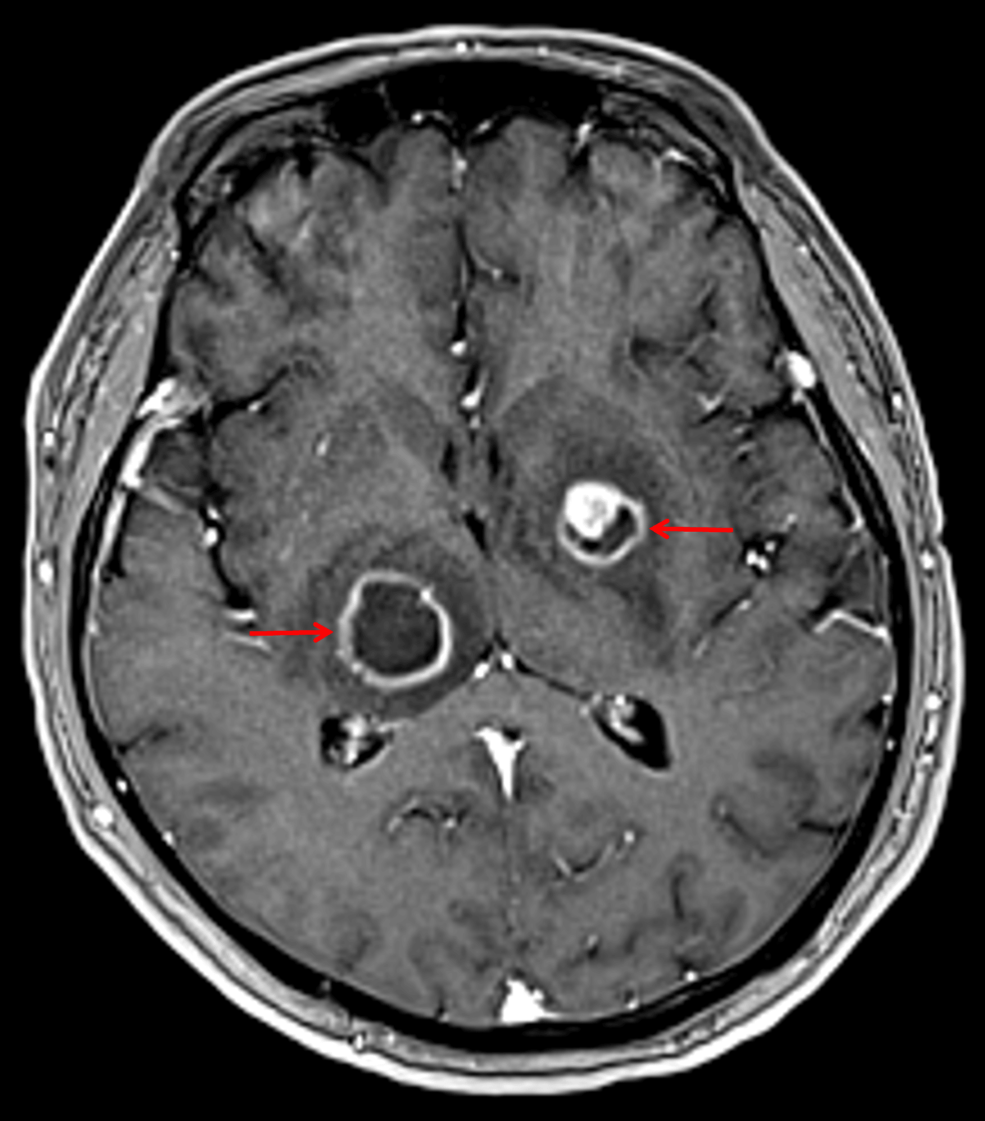
Peripherally-enhancing lesions in the right thalamus and left lentiform nucleus (red arrows).
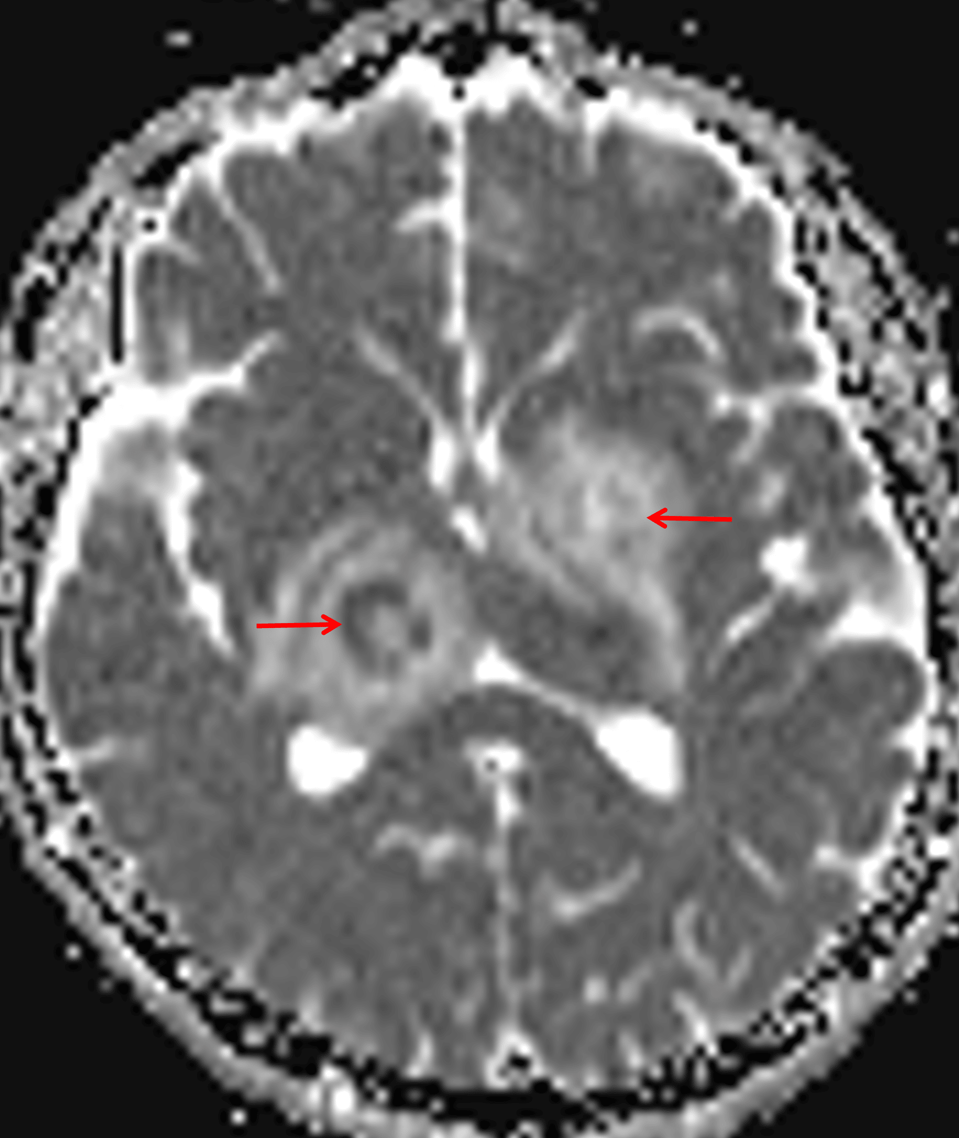
Peripheral restricted diffusion, more pronounced with the right thalamic lesion (red arrows).
Diagnosis
CNS toxoplasmosis
Key Imaging Features
Become a PRO member to unlock the key imaging features
Differential Diagnosis
Become a PRO member to unlock the differential diagnosis
Discussion
Pearls
Become a PRO member to unlock the pearls