Demographics:
21 years old, Female
Indication:
Transfer for hydrocephalus
Findings
CT with contrast
- Mildly hyperattenuating mass in the body of the right lateral ventricle with cystic components along its medial margin and broad contact with the septum pellucidum
- Patchy internal areas of enhancement
MRI
- Heterogeneous mass in the body of the right lateral ventricle measuring 3.8 x 3.4 x 3.8 cm with a solid component laterally and multiple cystic components medially, broadly contacting the septum pellucidum
- The solid component is isointense on T2, mildly hyperintense on FLAIR, and demonstrates diffuse restricted diffusion and mild patchy enhancement
- Associated right lateral deviation of the septum pellucidum and mass effect on the third ventricle with resultant obstructive hydrocephalus involving the lateral ventricles and anterior aspect of the third ventricle
Annotated Images & Illustrations
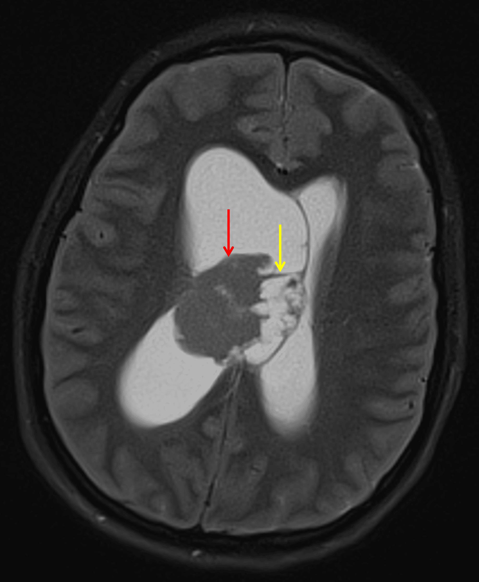
Mass in the body of the right lateral ventricle with a solid component laterally (red arrow) and a cystic component medially (yellow arrow).
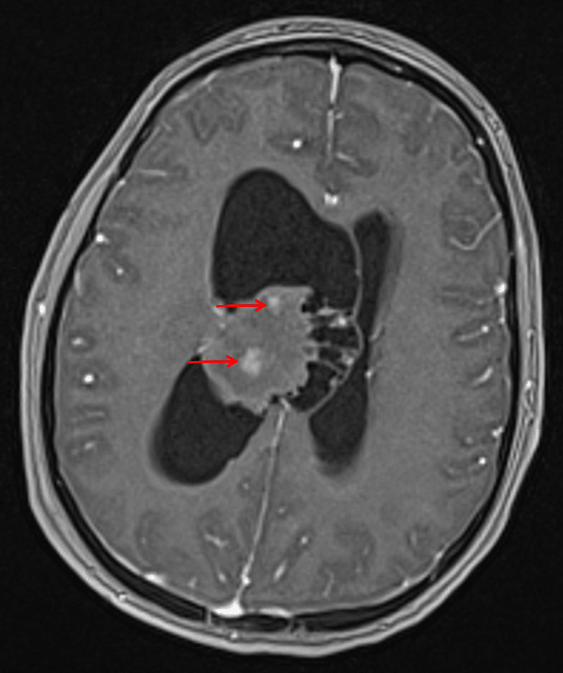
Internal patchy areas of enhancement (red arrows).
Diagnosis
Central neurocytoma
Differential Diagnosis
Become a PRO member to unlock the differential diagnosis
Pearls
Become a PRO member to unlock the pearls