Findings
CT
- Cystic mass in the left cerebellar hemisphere with a solid mural nodule posterosuperiorly
- No corresponding calcification
- Associated mass effect on the fourth ventricle and obstructive hydrocephalus with subependymal edema
MRI
- Cystic mass in the left cerebellar hemisphere with an enhancing mural nodule along its posterosuperior margin abutting the left tentorial leaflet
- Susceptibility artifact in the mural nodule, which may relate to internal vascularity
- No corresponding restricted diffusion
- Minimal rim of surrounding parenchymal edema
- Associated mass effect on the fourth ventricle and obstructive hydrocephalus with subependymal edema
Annotated Images & Illustrations
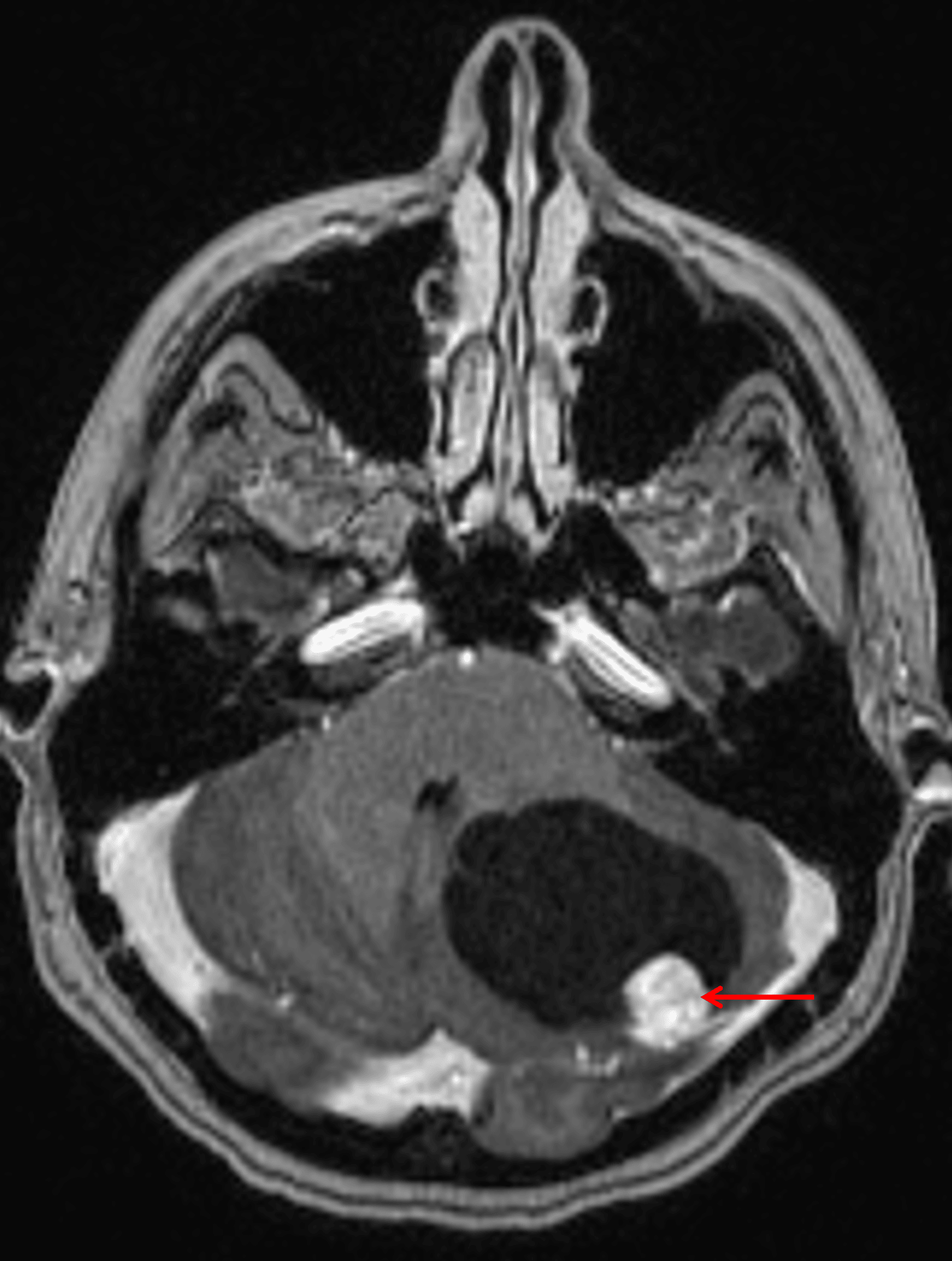
Cystic mass in the left cerebellar hemisphere with an avidly enhancing mural nodule (red arrow).
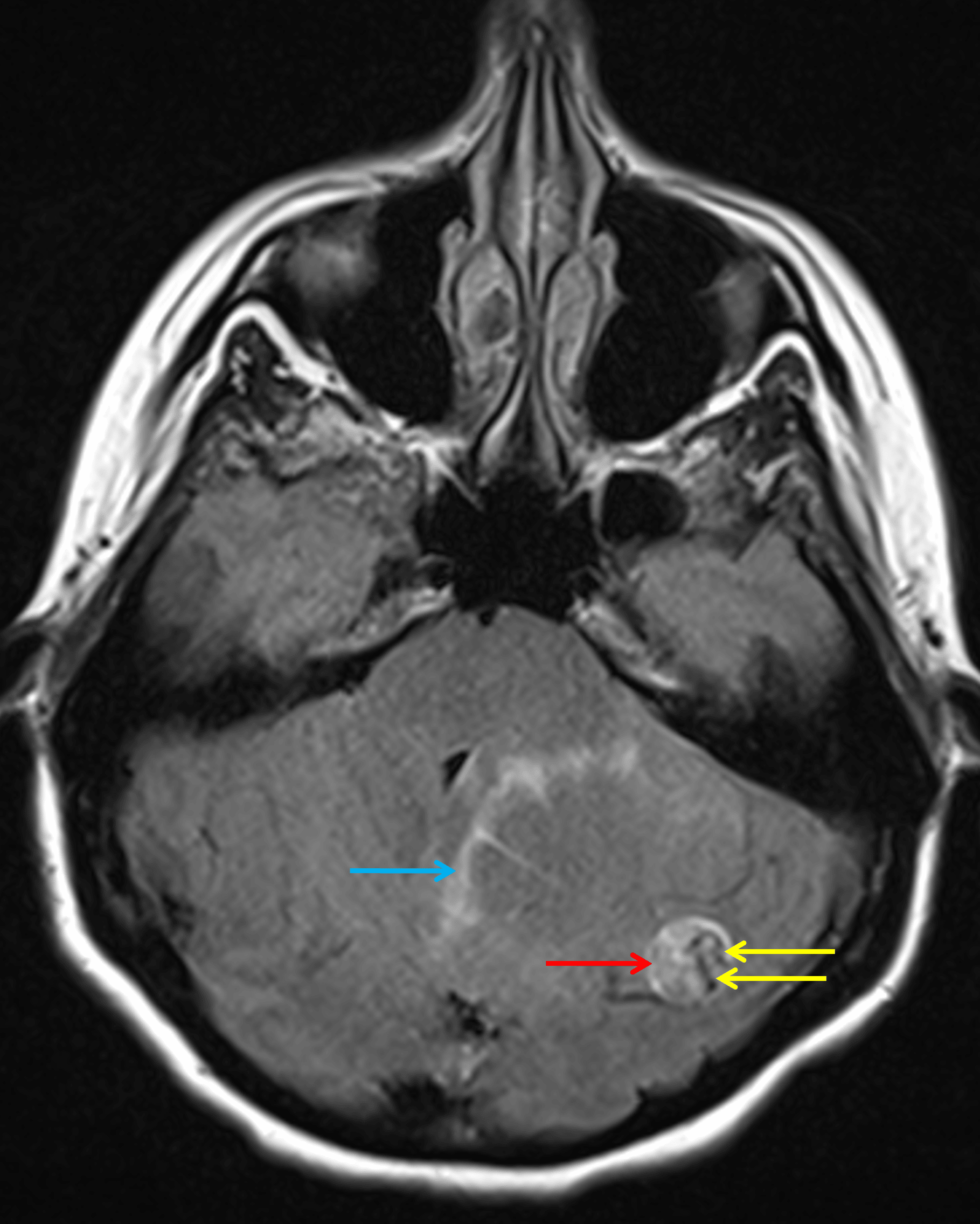
Axial FLAIR sequence demonstrates hyperintense signal within the solid component (red arrow) contrasted with serpiginous hypointense flow voids (yellow arrows). Minimal parenchymal edema is noted along the margins of the cystic component of the mass (blue arrow).
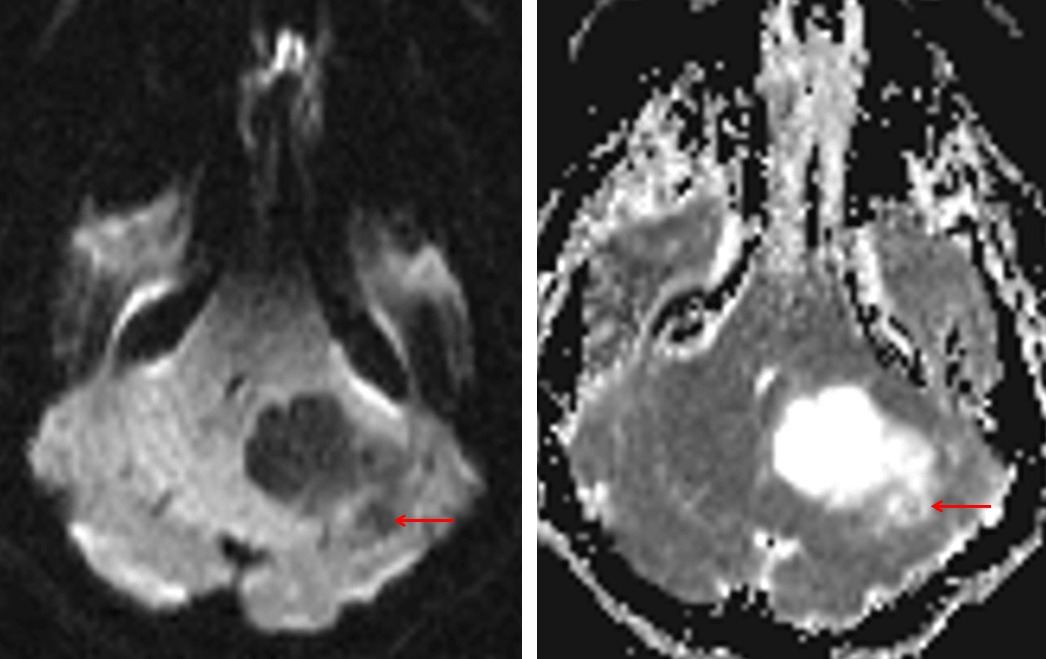
Relative facilitated diffusion (low DWI signal and high ADC signal, red arrows) is demonstrated in the solid portion of the mass relative to brain parenchyma.
Diagnosis
Hemangioblastoma
Key Imaging Features
Become a PRO member to unlock the key imaging features
Differential Diagnosis
Become a PRO member to unlock the differential diagnosis
Discussion
Pearls
Become a PRO member to unlock the pearls