Findings
CT
- Mixed cystic and solid suprasellar mass with internal coarse calcification
- Left frontal approach ventricular shunt catheter terminating in the frontal horn of the left lateral ventricle
- Asymmetric prominence of the frontal horn of the right lateral ventricle, which may represent differential shunting
MRI
- Mixed cystic and solid suprasellar mass measuring 3.5 x 2.3 x 4.2 cm with solid enhancement of the solid components and peripheral enhancement of the cystic components
- Intrinsic T1 signal hyperintensity within a few of the cystic components (particularly the most anterosuperior cyst), which may represent intralesional hemorrhage or proteinaceous debris
- The mass contacts and anteriorly displaces the optic chiasm and splays the optic tracts
- The pituitary gland is seen separate from the mass
Annotated Images & Illustrations
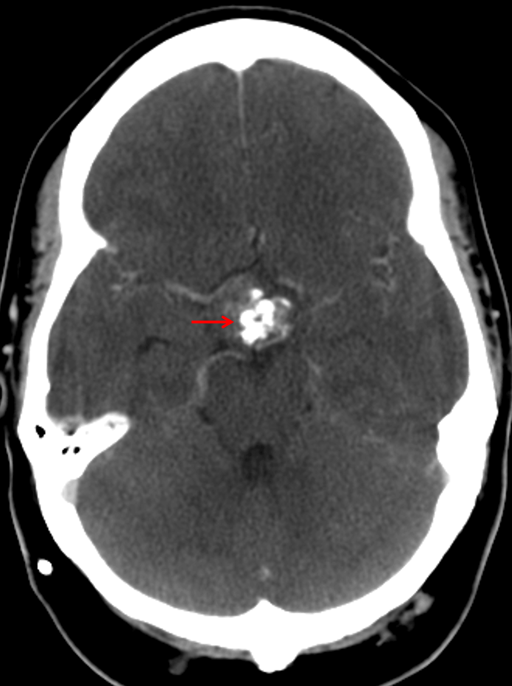
Suprasellar mass with coarse internal calcification (red arrow).
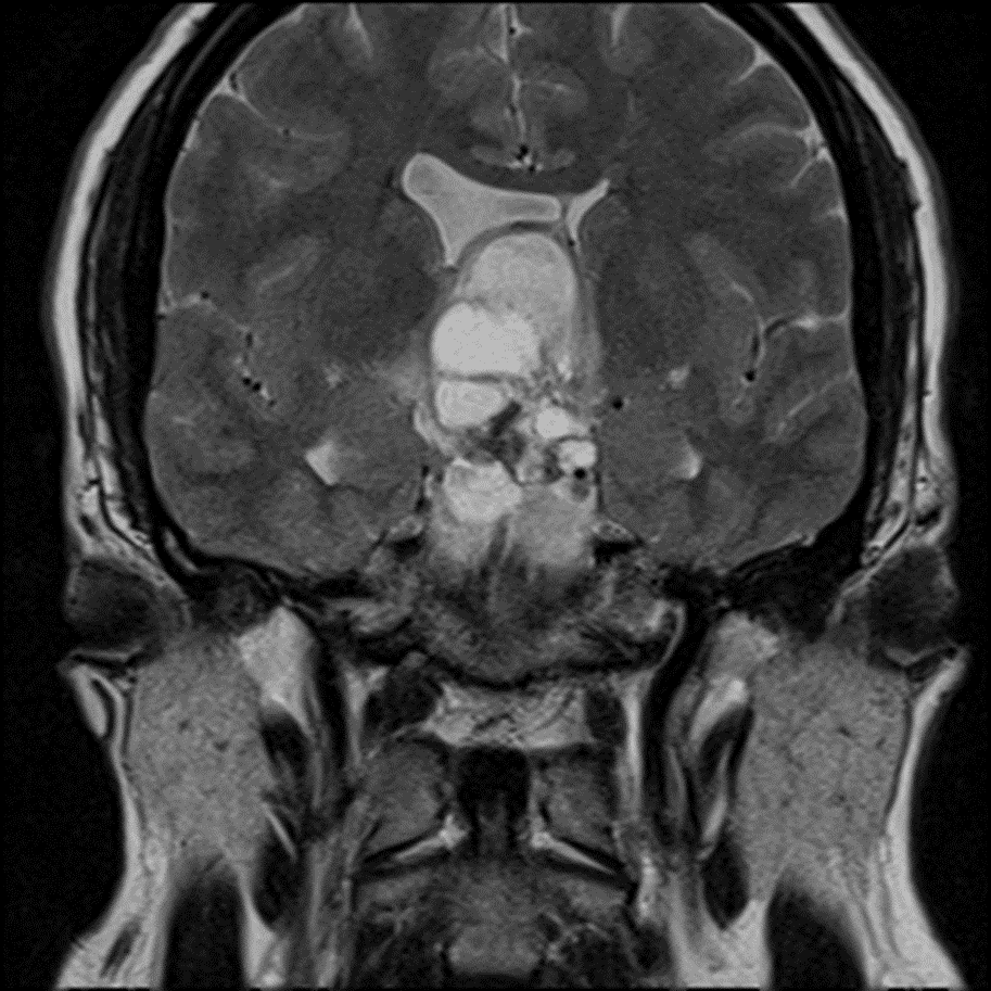
Multiple T2 hyperintense cystic components.
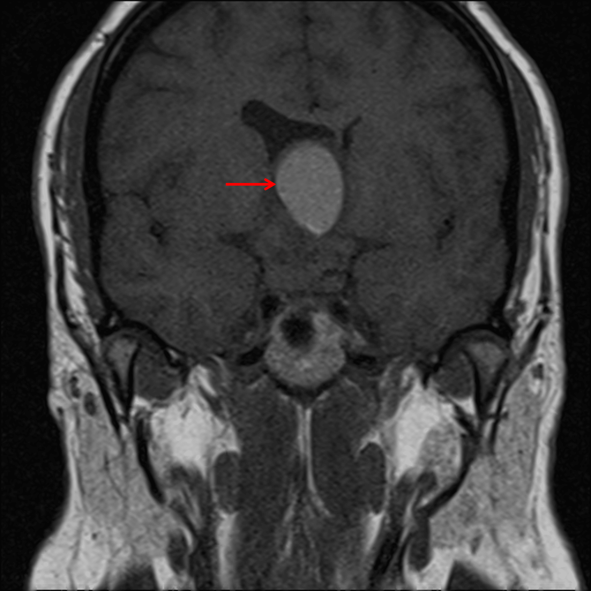
Intrinsic T1 signal hyperintensity within one of the larger superior cysts (red arrow).
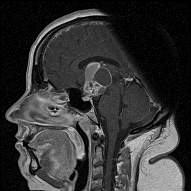
Solid enhancement of the solid components and peripheral enhancement of the cystic components.
Diagnosis
Adamantinomatous craniopharyngioma
Key Imaging Features
Become a PRO member to unlock the key imaging features
Differential Diagnosis
Become a PRO member to unlock the differential diagnosis
Discussion
Pearls
Become a PRO member to unlock the pearls