Demographics:
74 years old, Female
Indication:
Altered mental status
Findings
CT
- Areas of hypoattenuation in the right greater than left lentiform nuclei, internal capsules, and thalami with extension into the bilateral cerebral white matter
MRI
- Relatively symmetric T2/FLAIR signal hyperintensity most pronounced in the supratentorial deep gray nuclei and deep white matter, including the internal capsules and corpus callosum, with associated curvilinear and nodular enhancement in a perivascular distribution
- Patchy corresponding restricted diffusion
- No midline shift or evidence of herniation or hydrocephalus
Annotated Images & Illustrations
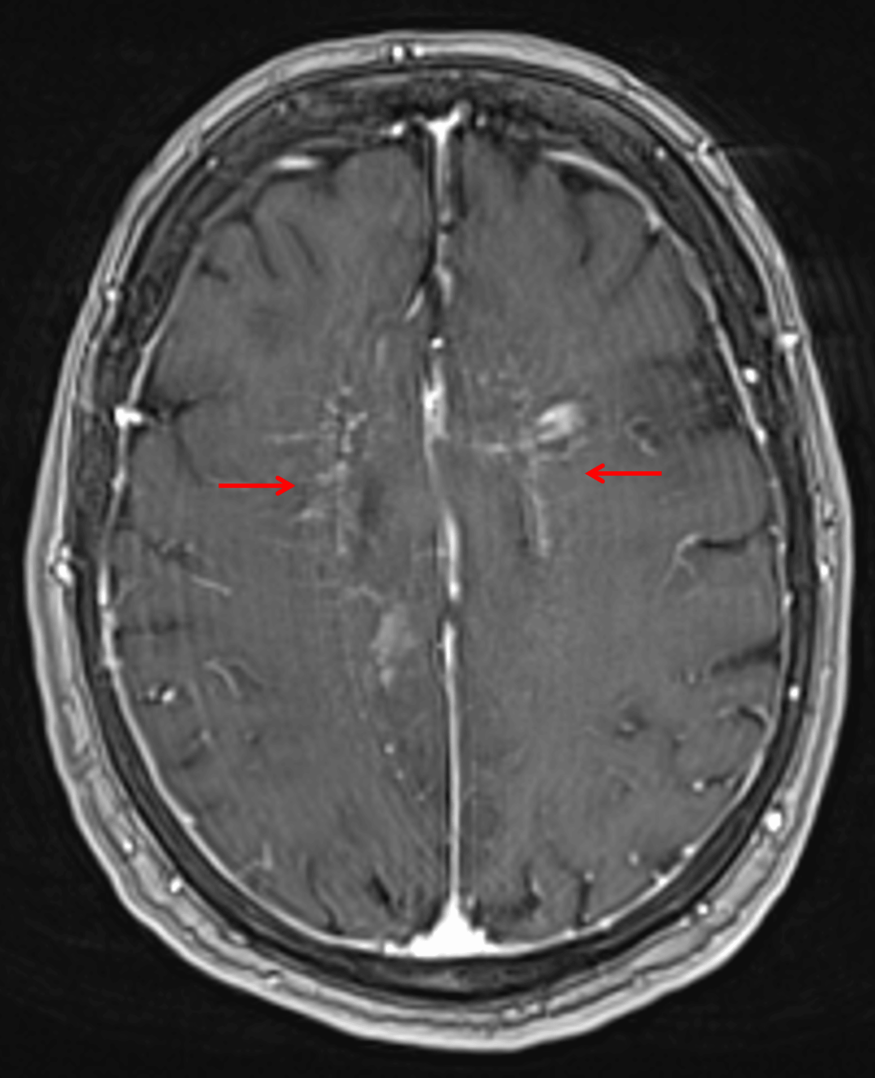
Curvilinear and nodular enhancement tracking along perivascular spaces in the bilateral cerebral white matter (red arrows) in this patient with intravascular lymphoma.
Diagnosis
Intravascular lymphoma
Key Imaging Features
Become a PRO member to unlock the key imaging features
Differential Diagnosis
Become a PRO member to unlock the differential diagnosis
Discussion
Pearls
Become a PRO member to unlock the pearls