Demographics:
68 years old, Female
Indication:
Confusion in setting of HIV/AIDS
Findings
- Multiple enhancing lesions in the bilateral deep gray nuclei and internal capsules, left hippocampal tail, subcortical right frontal lobe, subcortical posterior right temporal lobe, and along the lateral ependymal margin of the frontal horn of the right lateral ventricle
- Several lesions demonstrate central hypoenhancement
- Variable corresponding restricted diffusion
- Surrounding vasogenic edema and local mass effect without midline shift or evidence of herniation or hydrocephalus
- Asymmetric enhancement of the left mandibular nerve at and inferior to the foramen ovale
- Asymmetric edema and enhancement in the left masseter muscle and overlying soft tissues
- Presumed pituitary macroadenoma, measuring 13 mm in craniocaudal span, with partial encasement of the left cavernous internal carotid artery and contact with the optic chiasm
Annotated Images & Illustrations
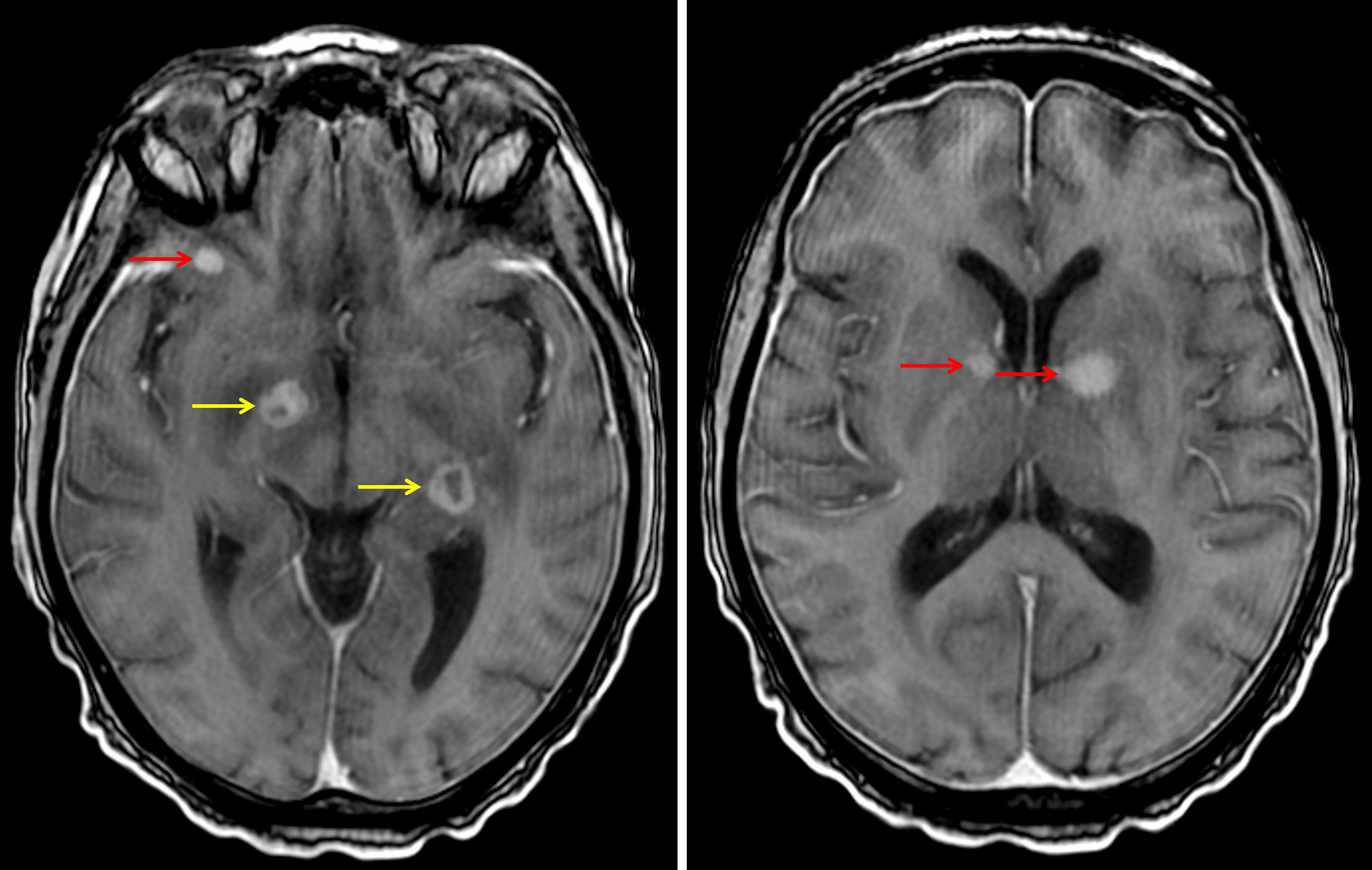
Multiple enhancing lesions intra-axial lesions with some demonstrating solid enhancement (red arrows) and others demonstrating peripheral enhancement (yellow arrows).
Diagnosis
Immunodeficiency-associated CNS lymphoma
Key Imaging Features
Become a PRO member to unlock the key imaging features
Differential Diagnosis
Become a PRO member to unlock the differential diagnosis
Discussion
Pearls
Become a PRO member to unlock the pearls