Demographics:
2 years old, Female
Indication:
Imbalance, large nevus on back
Findings
- Multiple T1 hyperintense lesions predominantly centered within the cortex with largest foci in the medial left temporal lobe/amygdala and the left hemipons
- Cavum veli interpositi
- No abnormal intracranial enhancement
- No substantial intracranial mass effect or hydrocephalus
Annotated Images & Illustrations
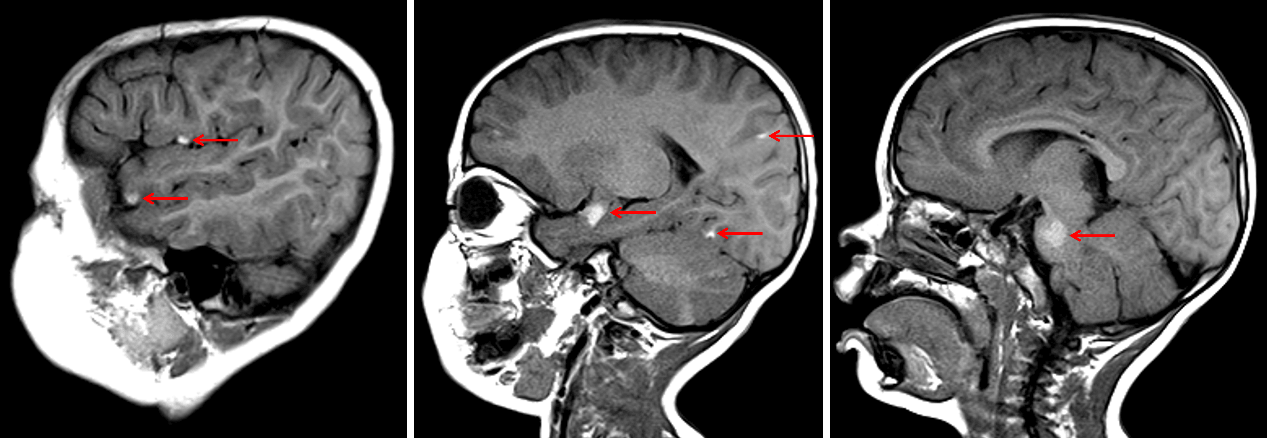
Multiple T1 hyperintense lesions scattered along the surface of the brain and brainstem (red arrows) in this patient with neurocutaneous melanosis.
Diagnosis
Meningeal melanocytosis (neurocutaneous melanosis)
Key Imaging Features
Become a PRO member to unlock the key imaging features
Differential Diagnosis
Become a PRO member to unlock the differential diagnosis
Discussion
Pearls
Become a PRO member to unlock the pearls