Demographics:
2 years old, Female
Indication:
Failure to thrive
Findings
CT (performed a few days after the MRI)
- Hypoattenuating suprasellar mass extending into the third ventricle with associated obstructive hydrocephalus and periventricular edema
- No calcification is identified within the mass
- Right frontal approach ventricular shunt catheter terminates in the third ventricle abutting the mass
MRI
- Avidly T2 hyperintense, homogeneously enhancing suprasellar mass which extends superiorly into the third ventricle, extends posteriorly into the prepontine cistern with mild mass effect on the pons, and encases the optic chiasm, bilateral supraclinoid internal carotid arteries, distal basilar artery, and the proximal portions of the bilateral posterior cerebral and superior cerebellar arteries
- No corresponding restricted diffusion or susceptibility artifact
- Associated obstructive hydrocephalus at the level of the third ventricle with periventricular edema
Annotated Images & Illustrations
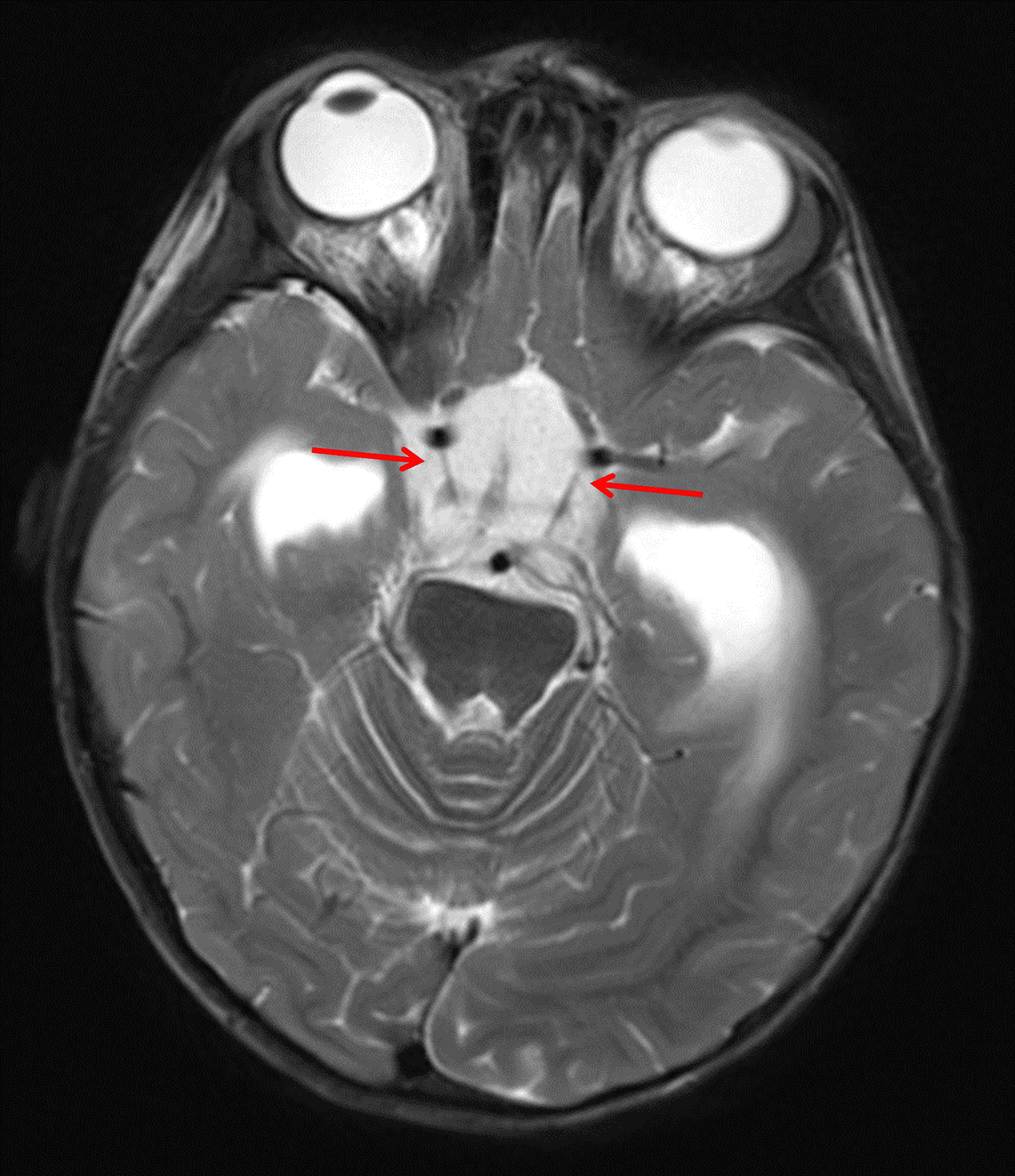
Markedly T2 hyperintense mass centered in the suprasellar cistern (red arrows).
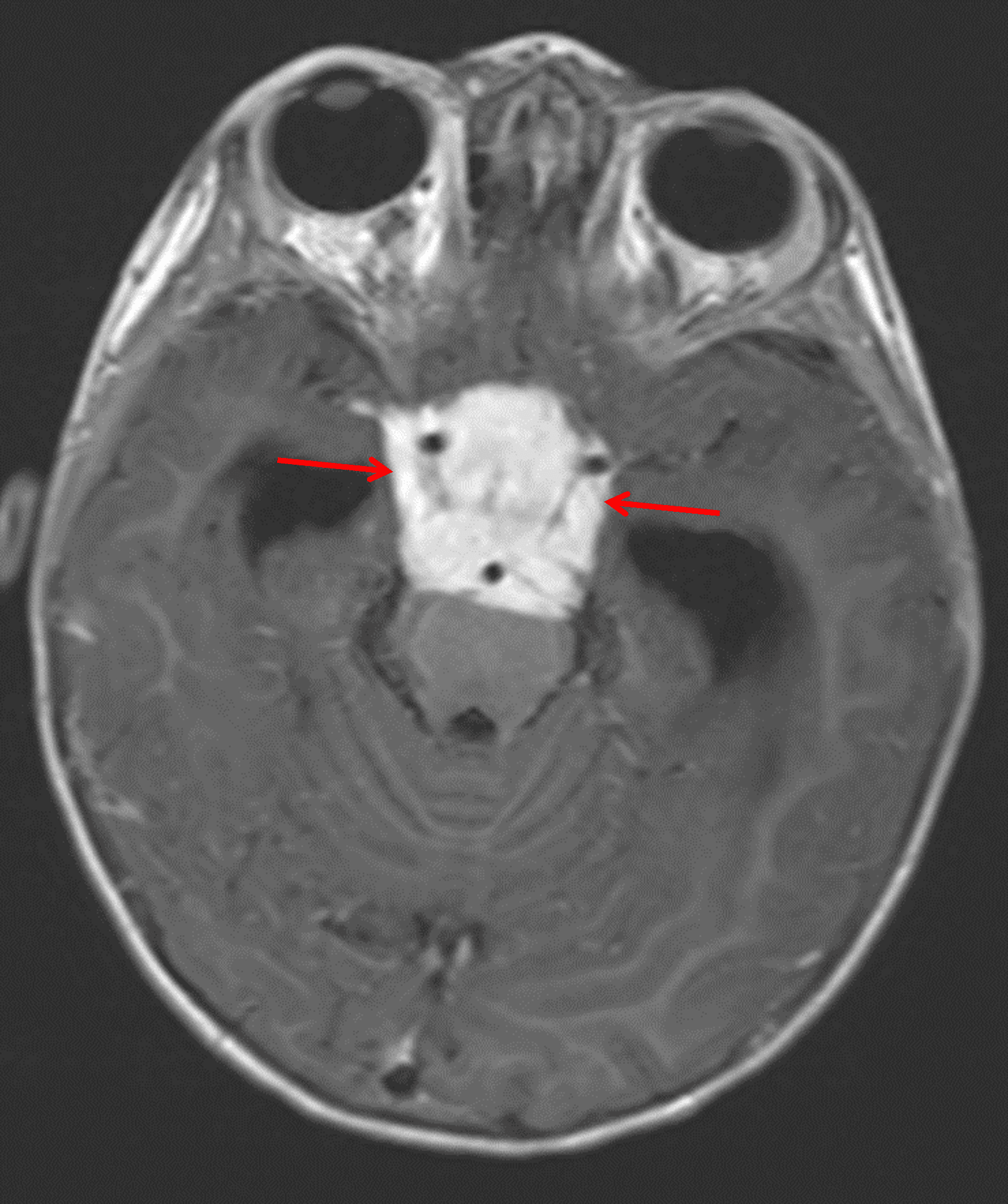
Corresponding avid enhancement (red arrows). The mass encases multiple vascular structures and extends into the prepontine cistern with mass effect on the pons.
Diagnosis
Pilomyxoid astrocytoma
Key Imaging Features
Become a PRO member to unlock the key imaging features
Differential Diagnosis
Become a PRO member to unlock the differential diagnosis
Discussion
Pearls
Become a PRO member to unlock the pearls