Findings
- Mixed solid and cystic pineal region mass measuring 23 x 20 x 15 mm
- The solid components diffusely enhance and demonstrate restricted diffusion
- Susceptibility artifact along the periphery of the mass, likely representing calcification
- Associated downward mass effect on the tectum with narrowing of the cerebral aqueduct and upstream hydrocephalus
Annotated Images & Illustrations
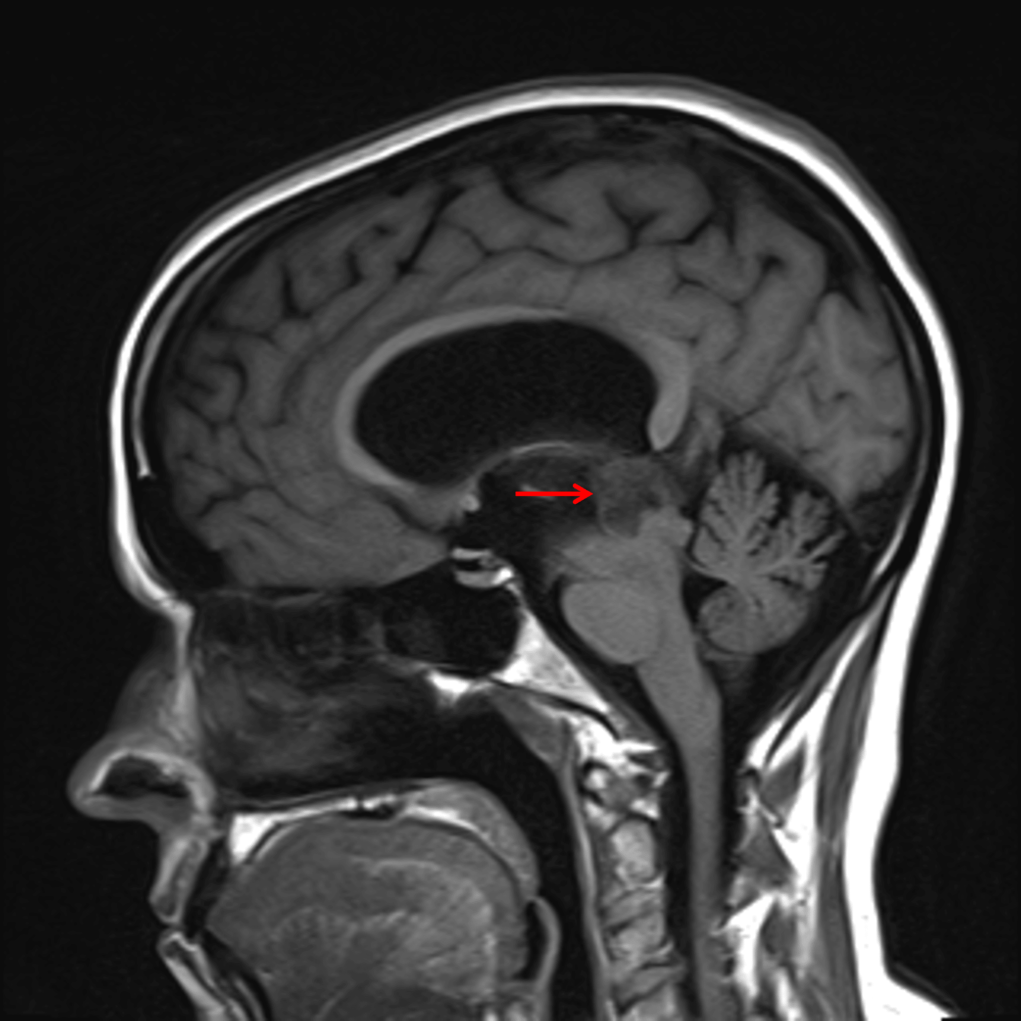
T1 hypointense pineal region mass (red arrow) with downward mass effect on the tectum and associated obstructive hydrocephalus.
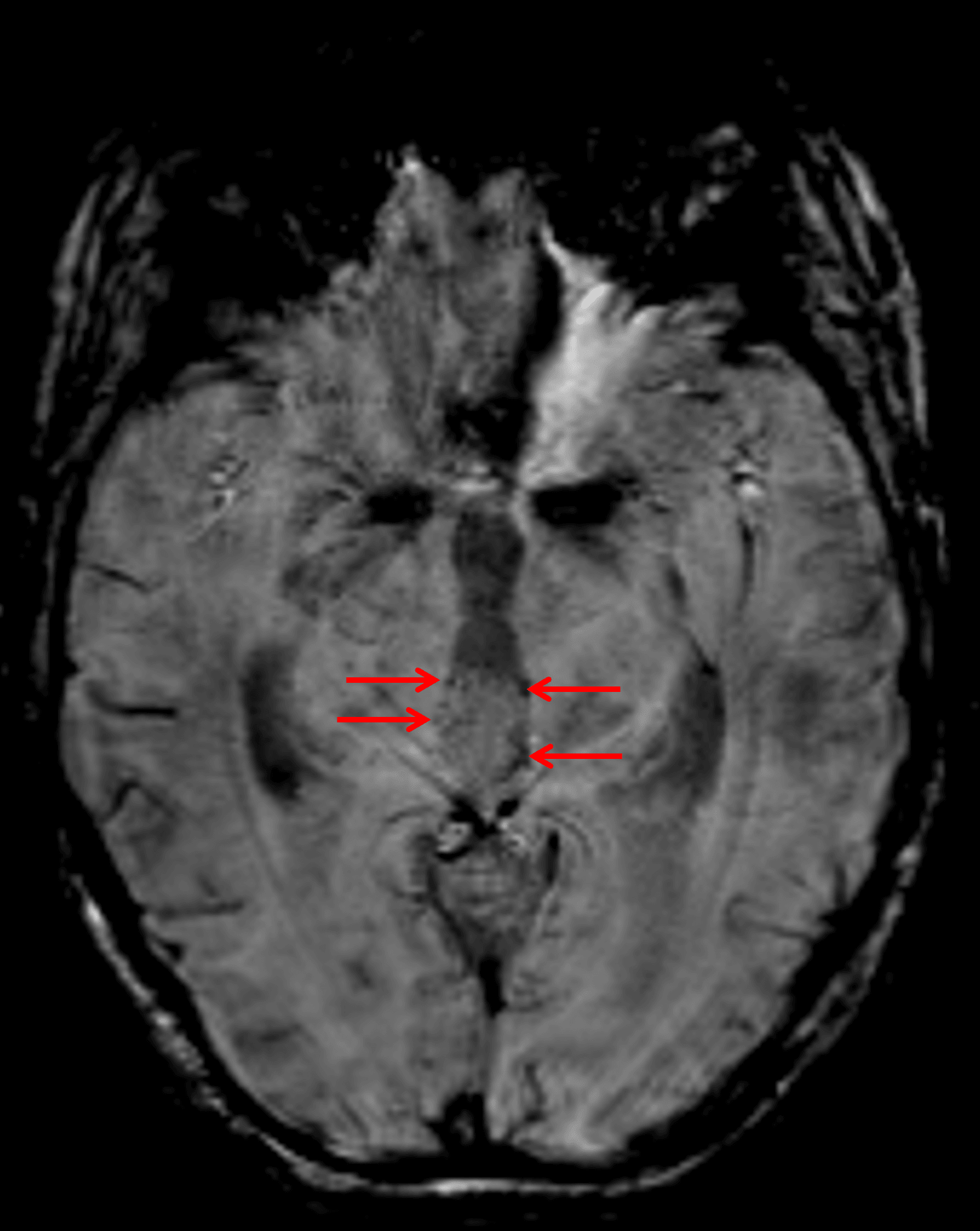
Foci of susceptibility artifact along the periphery of the mass (red arrows) represent peripheral calcification.
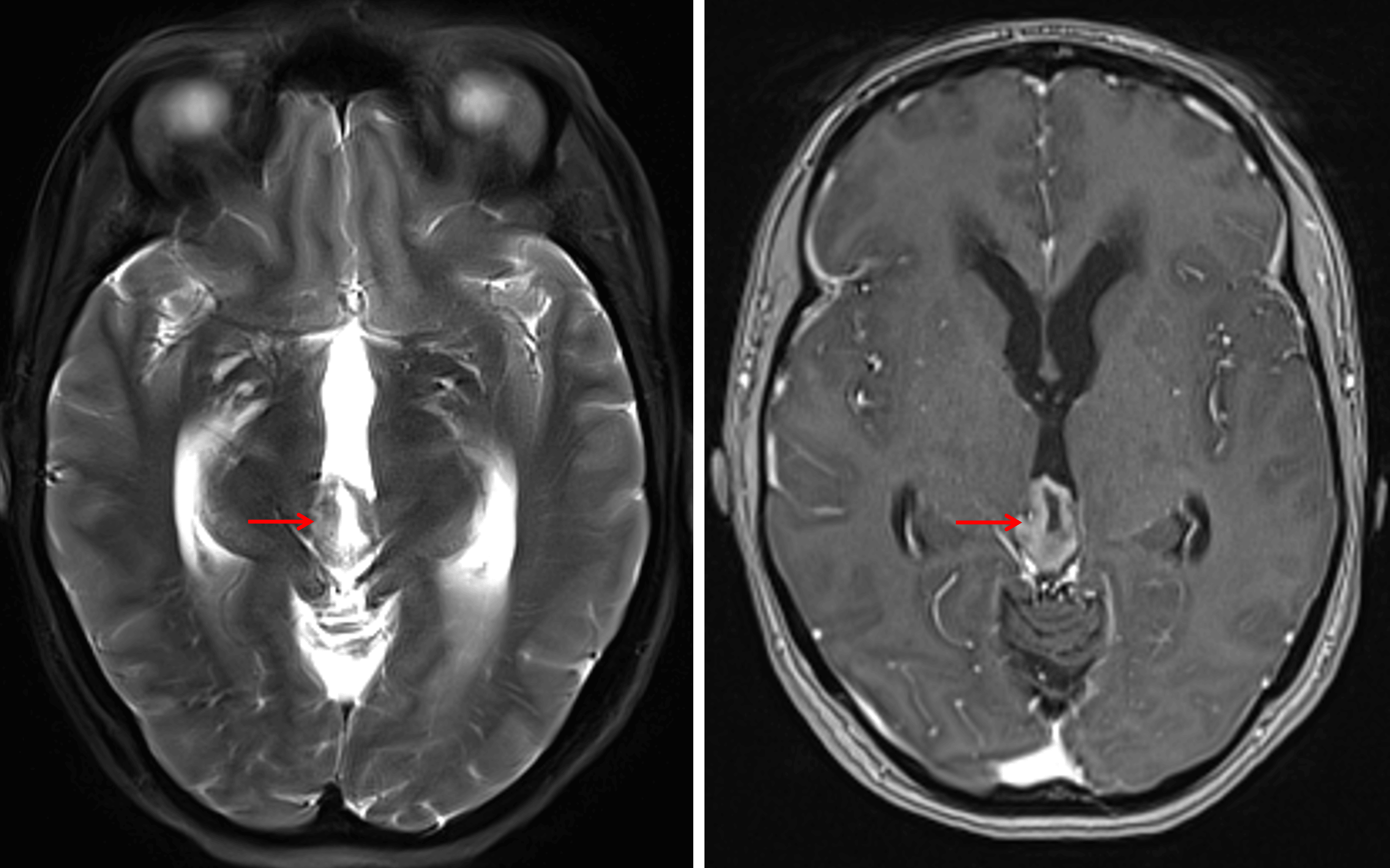
The mass has an internal cystic component with avid enhancement of the solid components (red arrows).
Diagnosis
Pineocytoma
Key Imaging Features
Become a PRO member to unlock the key imaging features
Differential Diagnosis
Become a PRO member to unlock the differential diagnosis
Discussion
Pearls
Become a PRO member to unlock the pearls