Demographics:
76 years old, Female
Indication:
Meningioma follow-up
Findings
- Heterogeneous, nonenhancing mass in the left cerebellar hemisphere which is mildly T2 hyperintense relative to the adjacent cerebellar parenchyma
- Mild diffuse diffusion signal hyperintensity (largely representing T2 shine-through)
- Associated mass effect on the inferior aspect of the fourth ventricle
- Right cerebellopontine angle meningioma
- Diffuse dural thickening and enhancement, likely related to chronic shunting
- Right parietal approach ventriculostomy catheter traverses the right lateral ventricle
Annotated Images & Illustrations
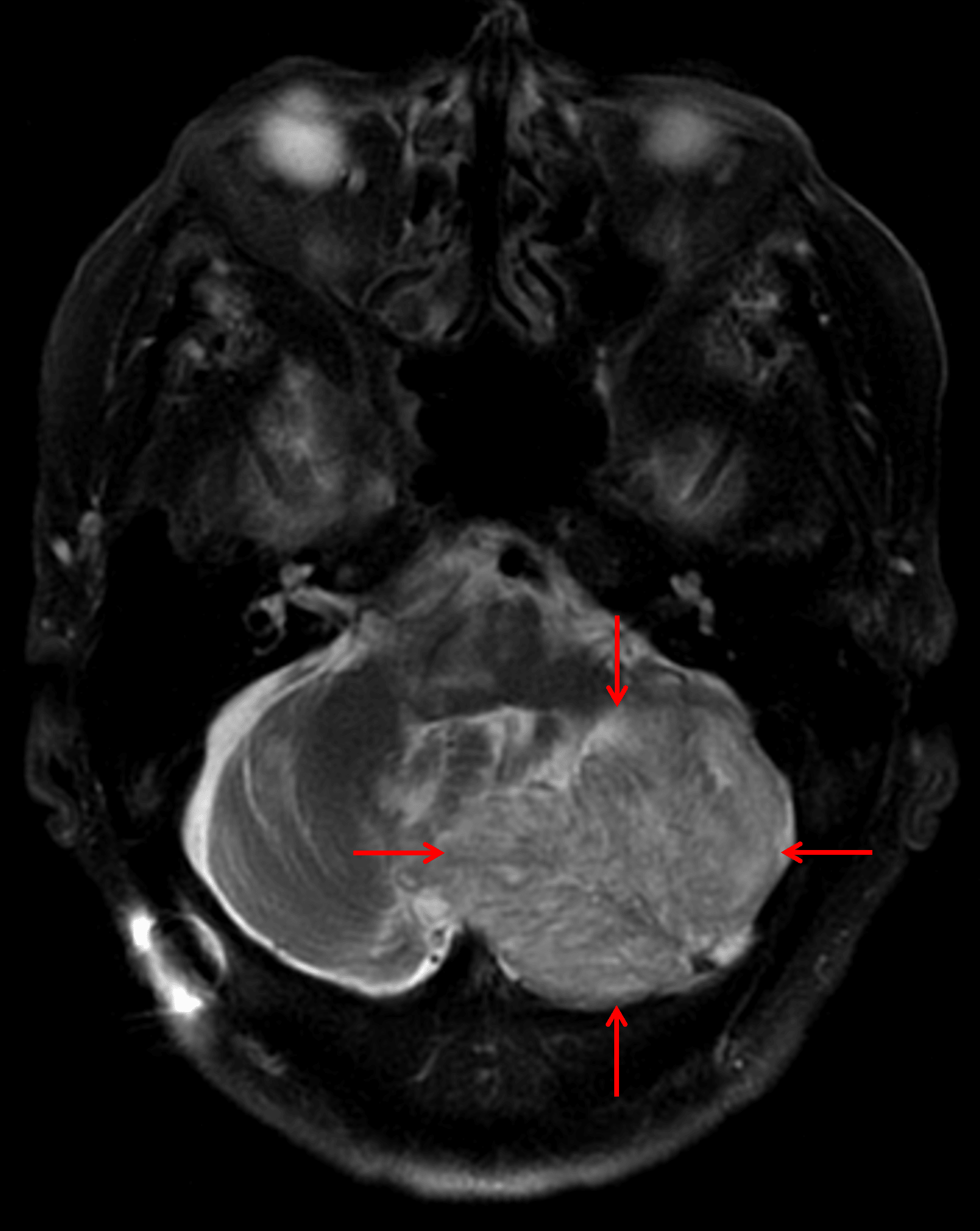
Mildly T2 hyperintense left cerebellar mass with striated internal architecture (red arrows), which is a classic appearance for a dysplastic cerebellar gangliocytoma.
Diagnosis
Dysplastic cerebellar gangliocytoma (Lhermitte Duclos disease)
Key Imaging Features
Become a PRO member to unlock the key imaging features
Differential Diagnosis
Become a PRO member to unlock the differential diagnosis
Discussion
Pearls
Become a PRO member to unlock the pearls