Demographics:
13 years old, Male
Indication:
Headaches
Findings
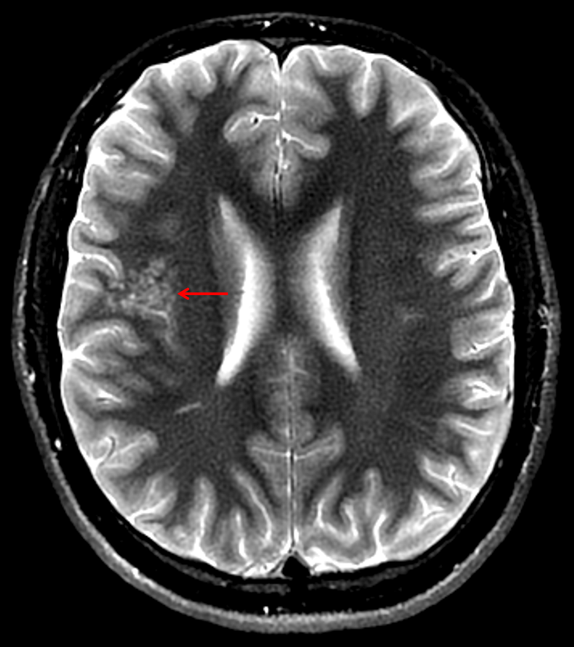
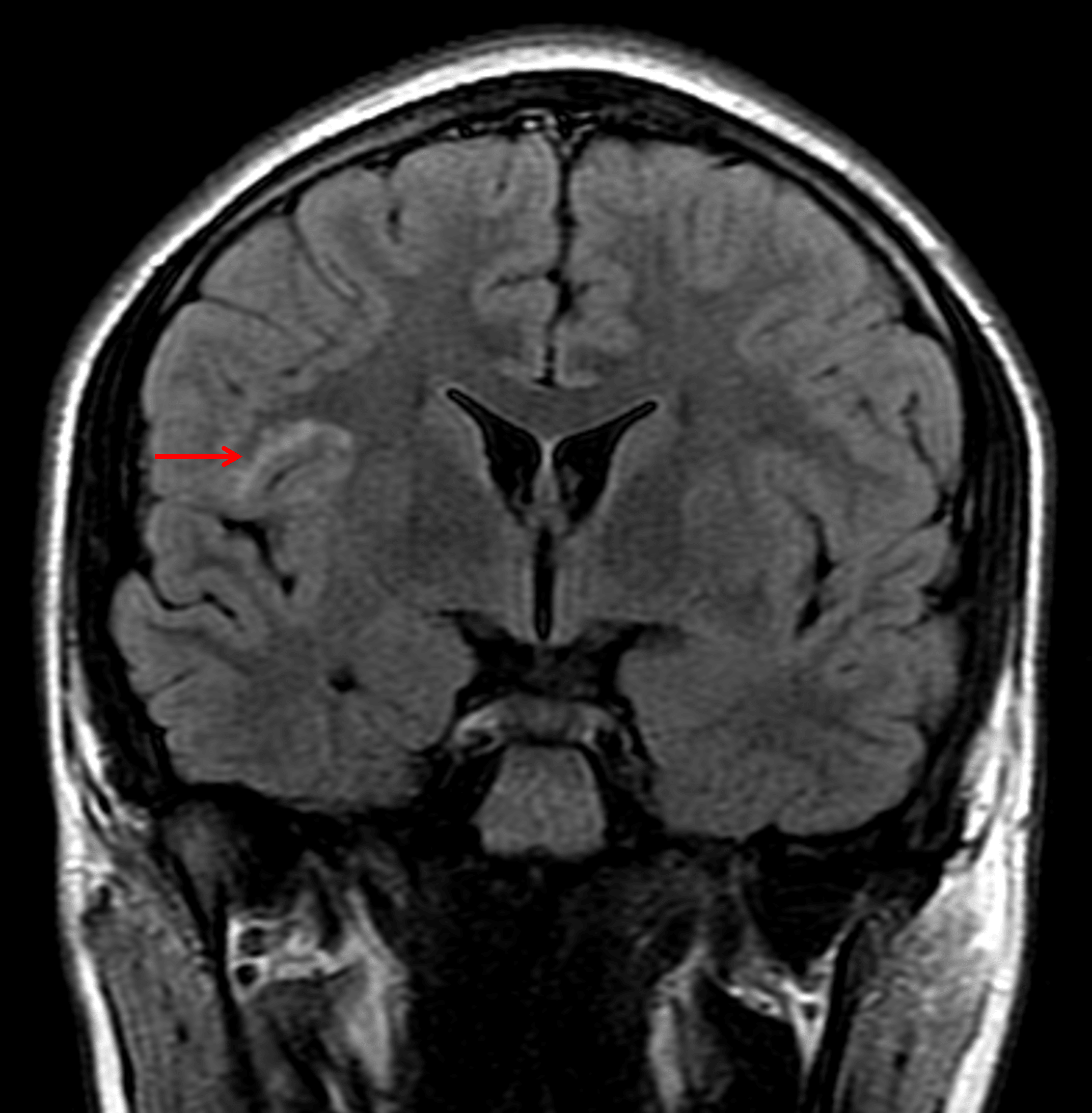
- Clustered T2/FLAIR hyperintense nodularity involving the right frontal operculum and superior insula
- Normal appearance of the overlying cortex
- No corresponding enhancement, restricted diffusion, or susceptibility artifact
Annotated Images & Illustrations
Clustered T2 hyperintense cystic structures in the subcortical white matter of the right frontal operculum and superior insula (red arrow) with normal appearance of the overlying cortex which is a typical appearance for MVNT.
This lesion remains hyperintense on FLAIR (red arrow).
Diagnosis
Multinodular and vacuolating neuronal tumor (MVNT)
Key Imaging Features
Become a PRO member to unlock the key imaging features
Differential Diagnosis
Become a PRO member to unlock the differential diagnosis
Discussion
Pearls
Become a PRO member to unlock the pearls