Demographics:
13 years old, Male
Indication:
Seizure
Findings
CT
- Wedge-shaped cortical and subcortical hypoattenuating mass in the left frontal lobe
MRI
- T2 hyperintense, cortically-based mass with lobulated margins in the left middle frontal gyrus measuring 20 x 18 x 20 mm
- Areas of internal FLAIR signal hypointensity
- No corresponding enhancement, restricted diffusion, or susceptibility artifact
- No surrounding edema or substantial mass effect
Annotated Images & Illustrations
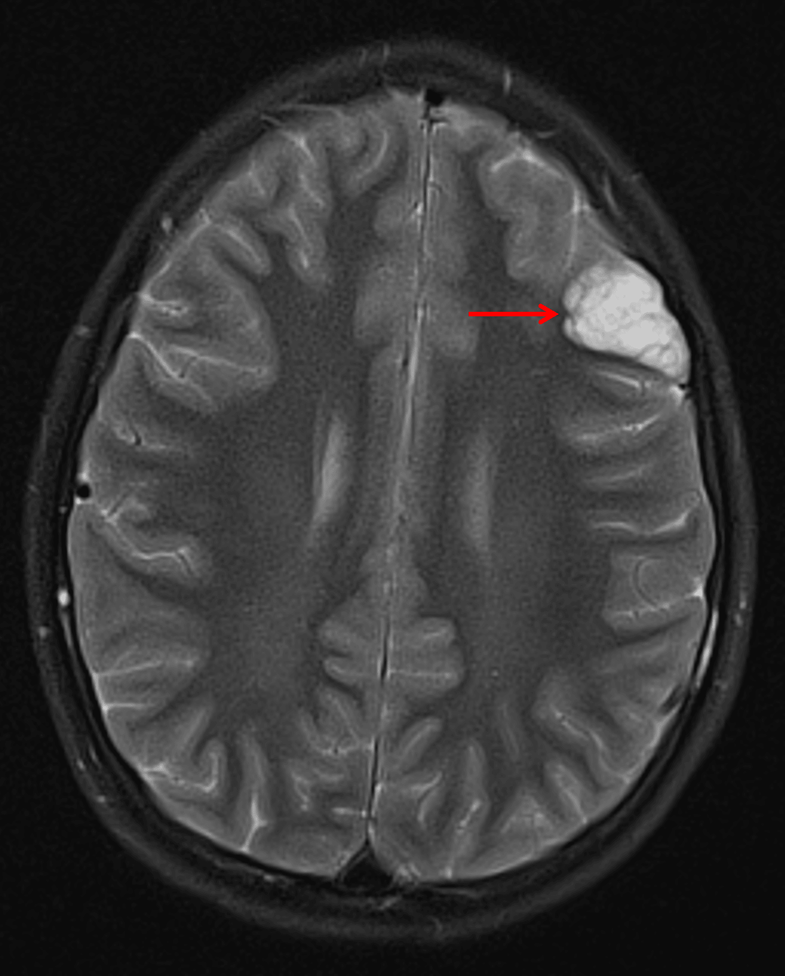
Wedge-shaped, cortically-based, bubbly T2 hyperintense lesion in the left frontal lobe (red arrow) consistent with a DNET.
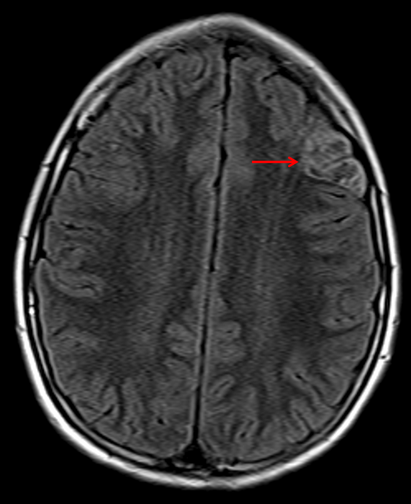
Note the heterogeneous internal FLAIR signal with a preserved hyperintense rim (red arrow).
Diagnosis
Dysembryoplastic neuroepithelial tumor (DNET)
Key Imaging Features
Become a PRO member to unlock the key imaging features
Differential Diagnosis
Become a PRO member to unlock the differential diagnosis
Discussion
Pearls
Become a PRO member to unlock the pearls