Demographics:
42 years old, Female
Indication:
Headache
Findings
- Intraventricular mass in the inferior aspect of the fourth ventricle measuring 12 x 10 x 10 mm
- No corresponding enhancement or restricted diffusion
- No hydrocephalus
- T2 hyperintense lesion adjacent to the frontal horn of the left lateral ventricle (patient had a history of multiple sclerosis)
- Dilated perivascular space in the right basal ganglia
Annotated Images & Illustrations
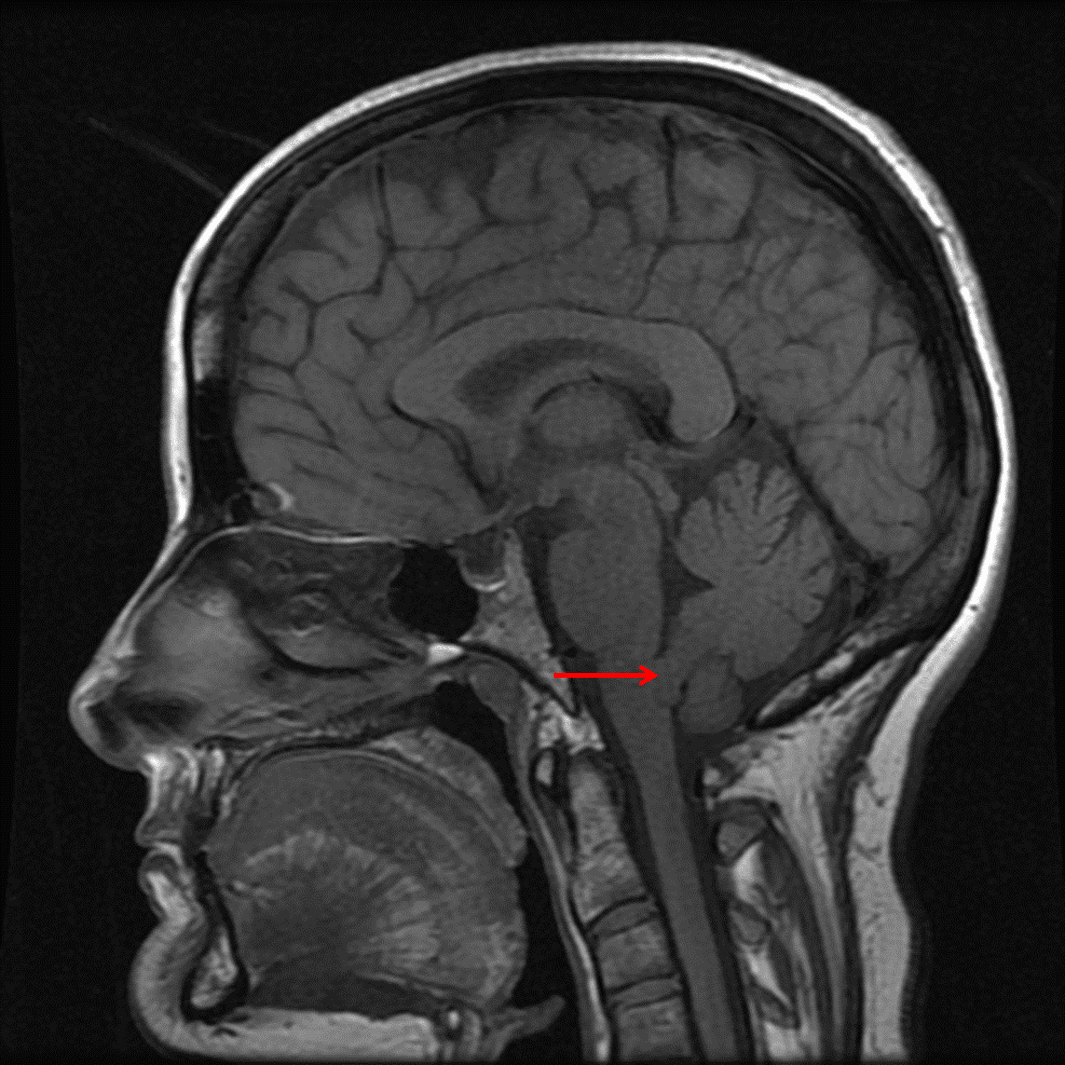
T1 isointense mass in the inferior aspect of the fourth ventricle (red arrow).
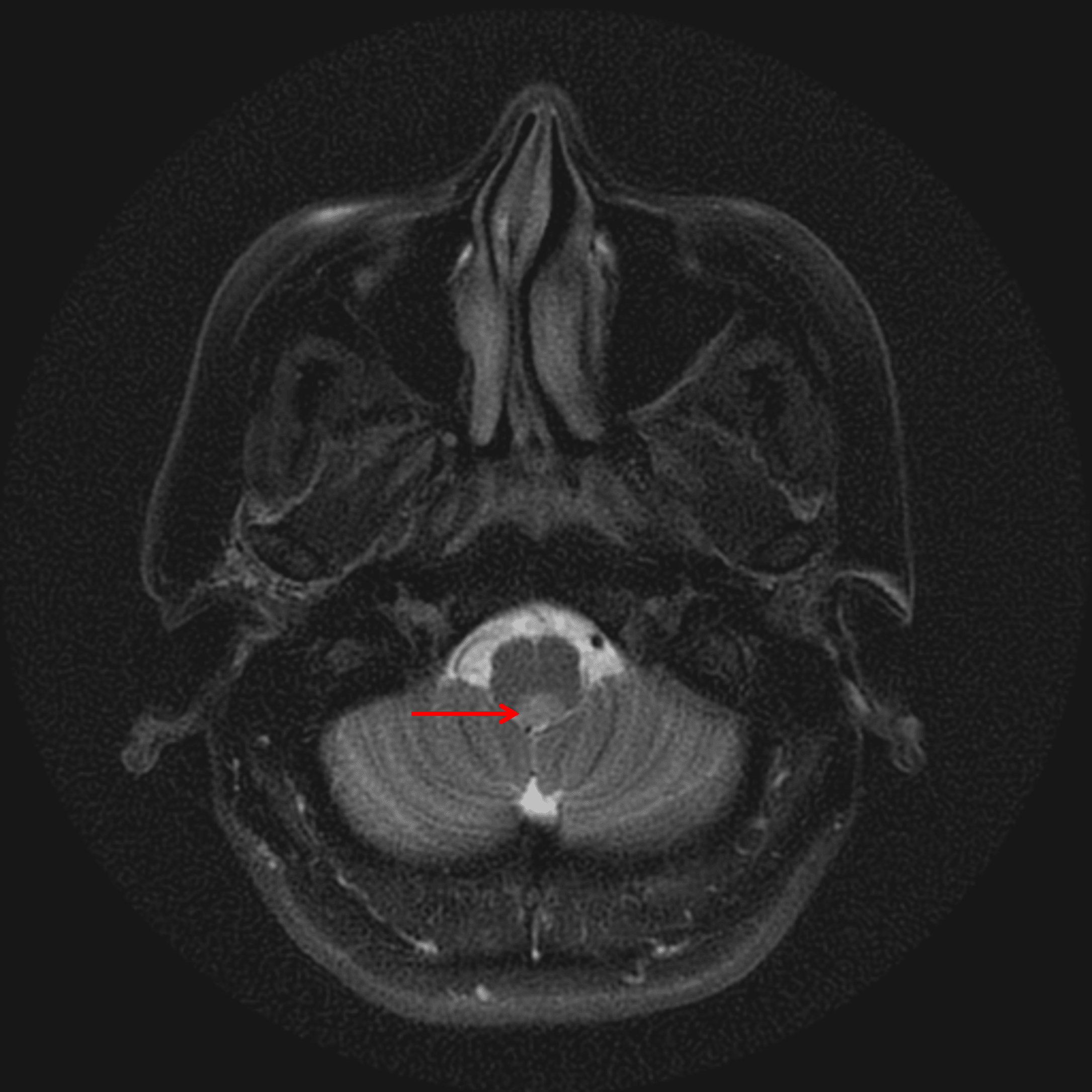
Mild corresponding T2 signal hyperintensity (red arrow).
Diagnosis
Subependymoma
Key Imaging Features
Become a PRO member to unlock the key imaging features
Differential Diagnosis
Become a PRO member to unlock the differential diagnosis
Discussion
Pearls
Become a PRO member to unlock the pearls