Utilisez la molette de la souris, les flèches ou le clic gauche (avec l'outil de défilement) pour faire défiler.
ui.case.use_touch_gestures
Aide DICOMSource: Local (us-east1-c)
1/28
Données démographiques:
2 ans, Féminin
Indication:
Failure to thrive
Constatations
CT (performed a few days after the MRI)
- Hypoattenuating suprasellar mass extending into the third ventricle with associated obstructive hydrocephalus and periventricular edema
- No calcification is identified within the mass
- Right frontal approach ventricular shunt catheter terminates in the third ventricle abutting the mass
MRI
- Avidly T2 hyperintense, homogeneously enhancing suprasellar mass which extends superiorly into the third ventricle, extends posteriorly into the prepontine cistern with mild mass effect on the pons, and encases the optic chiasm, bilateral supraclinoid internal carotid arteries, distal basilar artery, and the proximal portions of the bilateral posterior cerebral and superior cerebellar arteries
- No corresponding restricted diffusion or susceptibility artifact
- Associated obstructive hydrocephalus at the level of the third ventricle with periventricular edema
Images annotées et illustrations
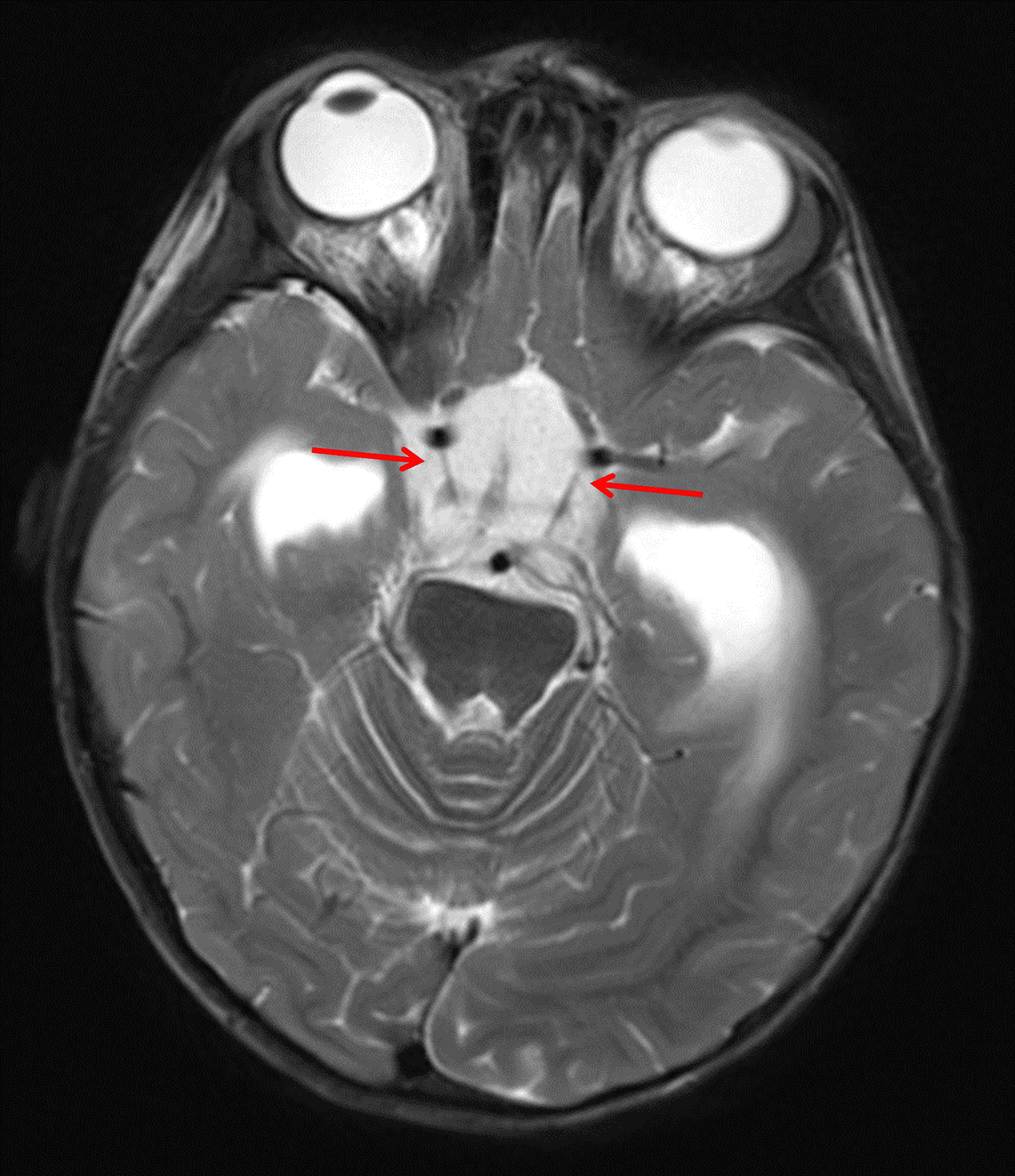
Markedly T2 hyperintense mass centered in the suprasellar cistern (red arrows).
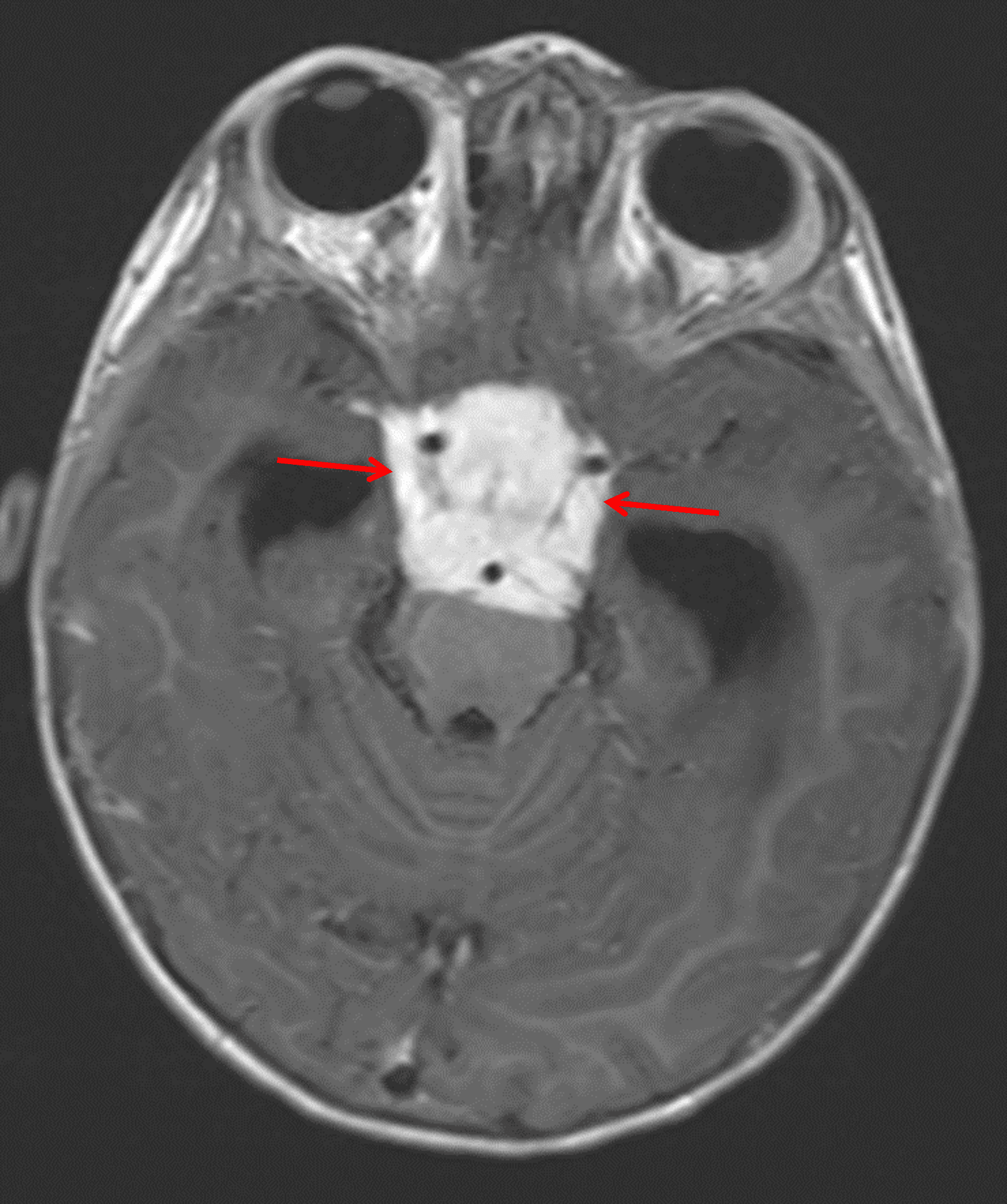
Corresponding avid enhancement (red arrows). The mass encases multiple vascular structures and extends into the prepontine cistern with mass effect on the pons.
Diagnostic
Pilomyxoid astrocytoma
Caractéristiques clés de l'imagerie
- These tumors have a strong predilection for the suprasellar region
- Due to myxoid components, these tumors are typically very T2 hyperintense and demonstrate facilitated rather than restricted diffusion
- Enhancement patterns are variable (when these tumors are not solidly enhancing and demonstrate internal hemorrhage, aneurysm is an important differential consideration)
- Internal hemorrhage is more common than with pilocytic astrocytomas
Diagnostic différentiel
- Pilocytic astrocytoma - Unlike PMAs, pilocytic astrocytomas are rare in children younger than 2 years old and are less likely to have internal hemorrhage
- Craniopharyngioma - Unlike PMAs, these commonly have calcification in children
- Germinoma - The suprasellar region is the second most common location for these avidly enhancing tumors (the pineal region is the most common location), but they tend to be less T2 hyperintense than PMAs and are much more common in male patients
Discussion
- Pilomyxoid astrocytomas (PMAs) occur across a large age range from early childhood to adulthood
- There is a potential association with neurofibromatosis type 1
Points essentiels
- Think about pilomyxoid astrocytoma when you see an avidly T2 hyperintense, enhancing suprasellar mass
- Compared to pilocytic astrocytomas, pilomyxoid astrocytomas are more likely to occur in very young children and are more likely to have associated hemorrhage