Utilisez la molette de la souris, les flèches ou le clic gauche (avec l'outil de défilement) pour faire défiler.
ui.case.use_touch_gestures
Aide DICOMSource: Local (us-east1-c)
1/25
Données démographiques:
7 ans, Masculin
Indication:
Screening MRI in patient with tuberous sclerosis
Constatations
- Interval enlargement of an enhancing subependymal nodule near the right foramen of Monro measuring 1.5 x 1 cm
- Multiple additional subcentimeter enhancing subependymal nodules
- Multiple T2/FLAIR hyperintense cortical and juxtacortical lesions in the bilateral cerebral hemispheres
- Dilated perivascular spaces in the right greater than left frontal white matter
- No hydrocephalus
- Right maxillary sinus mucous retention cyst. Mucosal thickening of bilateral ethmoid air cells
Images annotées et illustrations
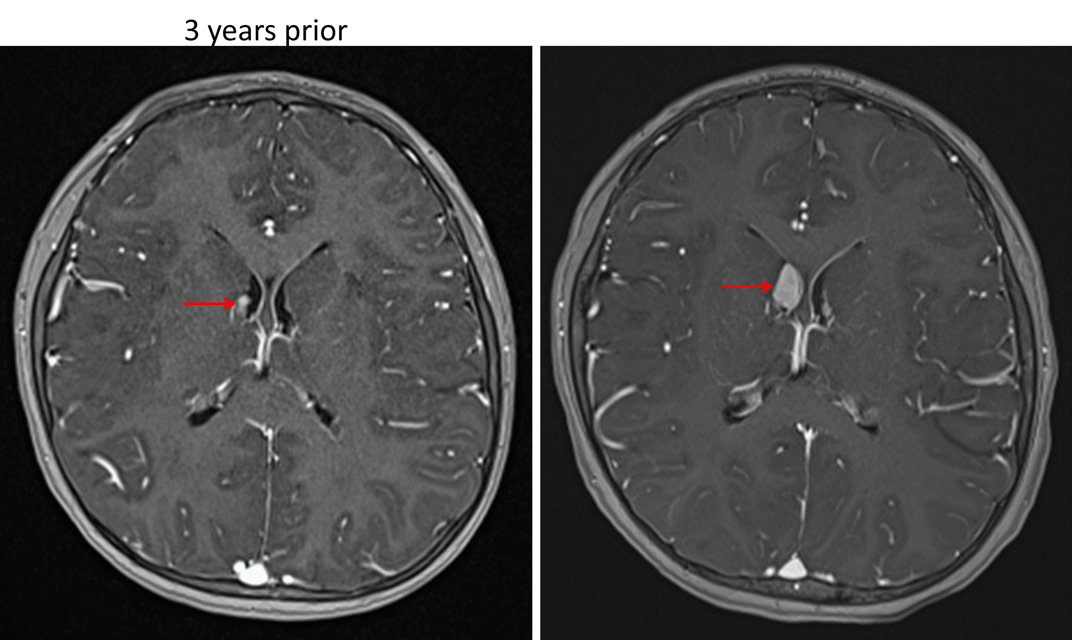
Enhancing subependymal nodule near the right foramen of Monro (red arrows) which demonstrates growth over a 3-year period, concerning for a subependymal giant cell astrocytoma in this patient with tuberous sclerosis.
Diagnostic
Subependymal giant cell astrocytoma (SEGA)
Caractéristiques clés de l'imagerie
- SEGAs are enhancing subependymal masses, commonly located near the foramen of Monro
- While SEGAs can be difficult to distinguish from subependymal nodules, growth over time and size exceeding 1 cm favor SEGA
Diagnostic différentiel
- No differential diagnosis in a patient with known tuberous sclerosis
Discussion
- Subependymal giant cell astrocytomas (SEGAs) are almost exclusively seen in patients with tuberous sclerosis
- They are thought to arise from subependymal nodules and typically present in later childhood or adolescence
- The primary concern with SEGAs is the development of hydrocephalus due to ventricular outflow obstruction at the foramen of Monro
Points essentiels
- In a patient with tuberous sclerosis, an enlarging subependymal nodule exceeding 1 cm in size near the foramen of Monro is highly concerning for a SEGA
- Watch closely for associated obstructive hydrocephalus