Utilisez la molette de la souris, les flèches ou le clic gauche (avec l'outil de défilement) pour faire défiler.
ui.case.use_touch_gestures
Aide DICOMSource: Local (us-east1-c)
1/36
Données démographiques:
5 ans, Féminin
Indication:
Headache, somnolence
Constatations
CT
- Hypoattenuating mass centered in the fourth ventricle with a more solid appearing component superiorly
- Associated obstructive hydrocephalus and periventricular edema
MRI
- Mixed solid and cystic mass centered in the fourth ventricle measuring 2.5 x 2.5 x 4 cm with a more solid enhancing component superiorly and a peripherally-enhancing cystic component inferiorly
- No corresponding restricted diffusion
- Associated effacement of the fourth ventricle and obstructive hydrocephalus with periventricular edema
Images annotées et illustrations
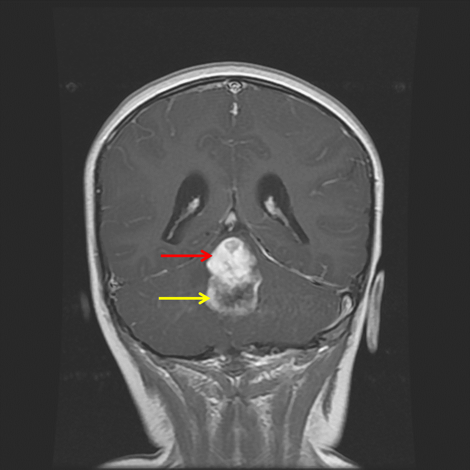
Midline posterior fossa mass with a superior solid component (red arrow) and inferior peripherally-enhancing cystic component (yellow arrow).
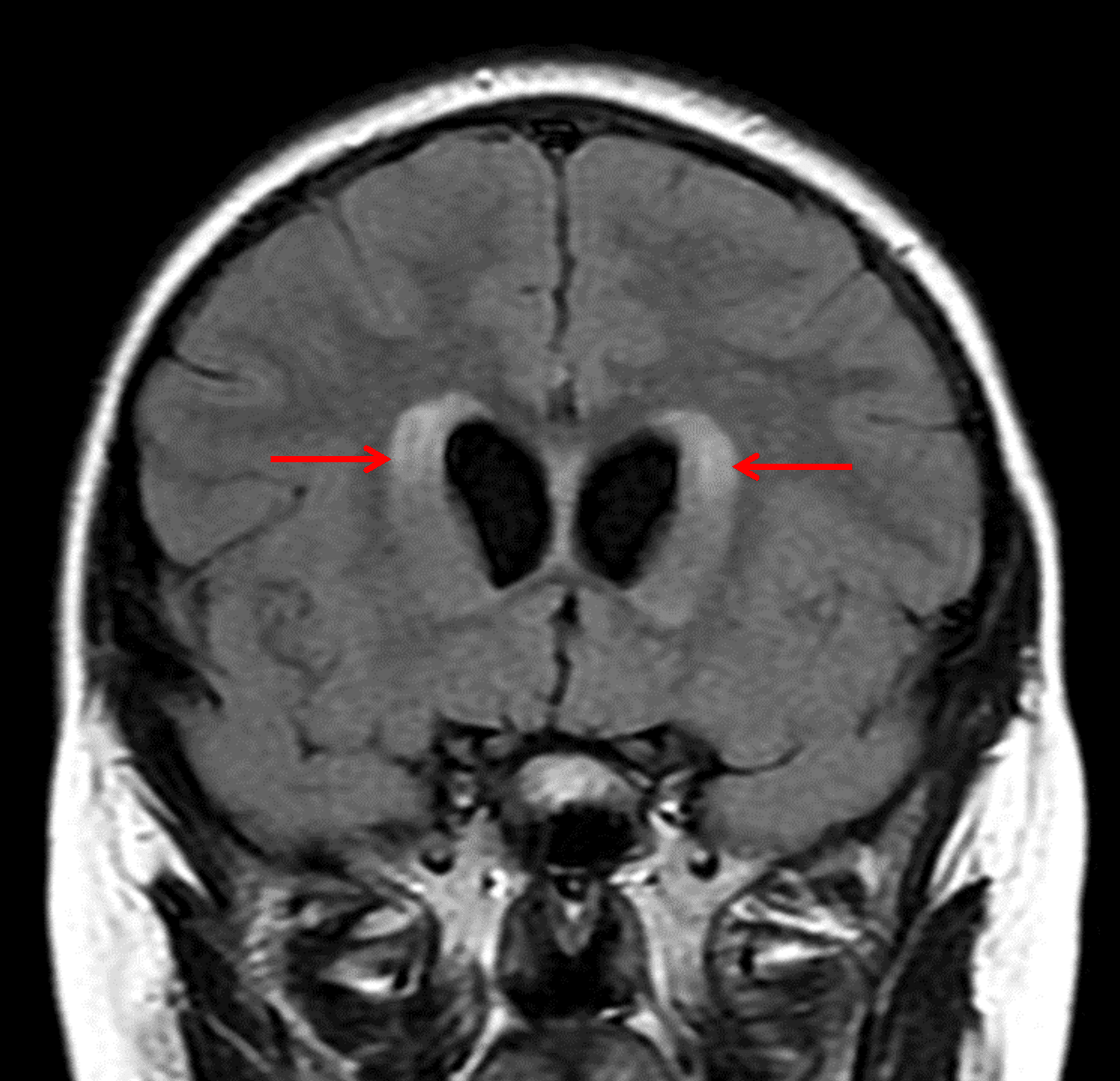
Associated obstructive hydrocephalus with subependymal edema (red arrows).
Diagnostic
Pilocytic astrocytoma
Caractéristiques clés de l'imagerie
- The "cyst with nodule" appearance is classic, but a minority of tumors do not demonstrate cystic components
- The solid component is usually hypoattenuating to brain on CT, T2 hyperintense, solidly enhancing, and does not demonstrate restricted diffusion
- Internal hemorrhage is rare
- Calcification is not usually seen
Diagnostic différentiel
- Ependymoma - Avidly enhancing tumors with a predilection for the fourth ventricle in children, these tend to be more malleable (including the characteristic appearance of tumor bulging through the foramina of Luschka) and are less likely to have cystic components
- Medulloblastoma - Avidly enhancing, commonly midline posterior fossa tumors, these tend to present at a younger age (3-9 years old) and classically have associated restricted diffusion
- Hemangioblastoma - Also "cyst with nodule" tumors, these are more common in adults and in patients with von Hippel Lindau disease
- High-grade glioma - These tend to occur in older children and are often more heterogeneous and poorly marginated than JPAs. Also, unlike JPAs, these have a propensity for CSF dissemination
Discussion
- Juvenile pilocytic astrocytomas (JPAs) are the most common posterior fossa tumor in children with a peak incidence between 5-13 years of age
- JPAs are WHO grade 1 tumors
- Most posterior fossa JPAs are sporadic; however optic pathway gliomas (OPGs) have a strong association with neurofibromatosis type 1
Points essentiels
- Think about JPA when you encounter a "cyst with nodule" mass, or any enhancing mass for that matter, in the posterior fossa in a child
- There is a strong association between optic pathway gliomas and NF1