Demographics:
5 years old, Female
Indication:
Headache, somnolence
Case #1
Findings
CT
- Hypoattenuating mass centered in the fourth ventricle with a more solid appearing component superiorly
- Associated obstructive hydrocephalus and periventricular edema
MRI
- Mixed solid and cystic mass centered in the fourth ventricle measuring 2.5 x 2.5 x 4 cm with a more solid enhancing component superiorly and a peripherally-enhancing cystic component inferiorly
- No corresponding restricted diffusion
- Associated effacement of the fourth ventricle and obstructive hydrocephalus with periventricular edema
Annotated Images & Illustrations
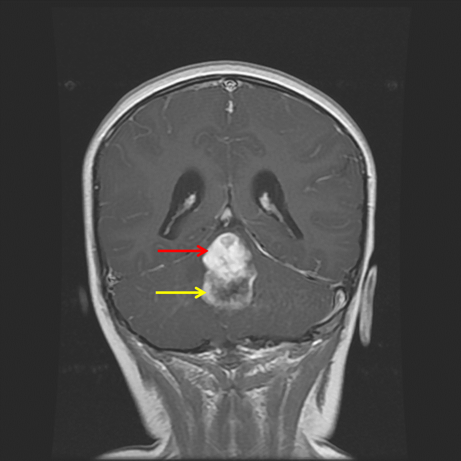
Midline posterior fossa mass with a superior solid component (red arrow) and inferior peripherally-enhancing cystic component (yellow arrow).
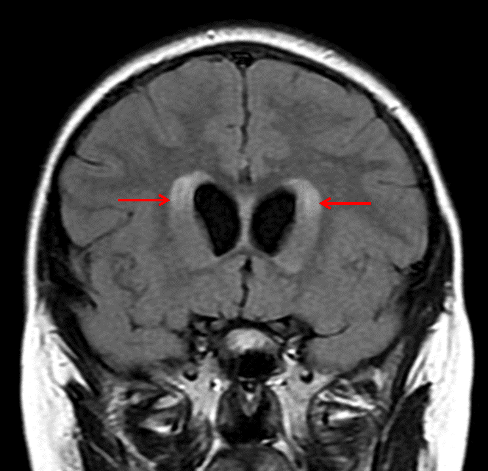
Associated obstructive hydrocephalus with subependymal edema (red arrows).
Diagnosis
Pilocytic astrocytoma
Key Imaging Features
Become a PRO member to unlock the key imaging features
Differential Diagnosis
Become a PRO member to unlock the differential diagnosis
Discussion
Pearls
Become a PRO member to unlock the pearls