Use mouse wheel, arrow keys or left click (with scroll tool selected) to scroll
Drag with one finger (with the scroll tool selected) to scroll
DICOM HelpSource: Local (us-east1-c)
Findings
-
No acute hemorrhage. Apparent thin extra-axial density overlying the high left frontal convexity on coronal imaging correlates with a normal draining cortical vein on the thin axial series.
- Note: This correlation was more apparent on PACS when using the localizer and scout line functionality.
Diagnosis
Cortical Vein Mimicking Hemorrhage
Discussion
-
Small draining cortical veins can mimic hemorrhage, particularly on the coronal reformat. The key to making the correct diagnosis is correlating with your thin axial sequence for the presence or absence of a correlate linear draining cortical vein.
-
If you are unsure if an extra-axial density represents hemorrhage or a small cortical vein it is reasonable to recommend short interval follow up CT.
-
Careful not to overlook a hyperdense thrombosed cortical vein, not an acute hemorrhage, but not just a benign vein!
-
Annotated Images & Illustrations
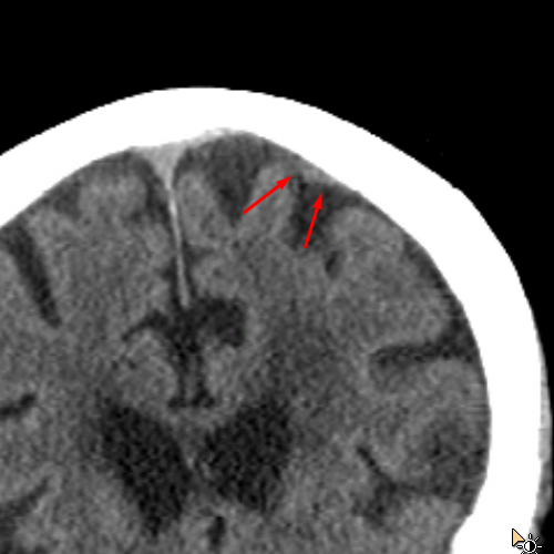
cortical-vein
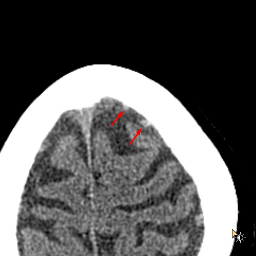
cortical-vein-axial
References