Use mouse wheel, arrow keys or left click (with scroll tool selected) to scroll
ui.case.use_touch_gestures
DICOM HelpSource: Local (us-east1-c)
Findings
- Acute oblique fracture of the distal fibula with approximately 1 cm override and posterolateral angulation
- Acute distracted fracture of the medial malleolus with approximately 2 cm lateral displacement
- Acute mildly comminuted fracture of the posterior malleolus with approximately 2 cm lateral displacement
- Posterolateral dislocation of the tibiotalar joint
- Marked widening of the distal tibiofibular syndesmosis
Diagnosis
Weber type C fracture
Ankle dislocation
Sample Report
Acute oblique fracture of the distal fibula with approximately 1 cm override and posterolateral angulation (Weber type C) with associated widening of the distal tibiofibular syndesmosis and posterior dislocation of the tibiotalar joint. MRI could further assess the extent of ligamentous injuries.
Acute distracted fracture of the medial malleolus with approximately 2 cm lateral displacement.
Acute mildly comminuted fracture of the posterior malleolus with approximately 2 cm lateral displacement.
Discussion
- While more complex ankle fracture classification systems exist, it is important to at least be familiar with the Weber classification:
- Weber type A: lateral malleolar fracture below the level of the talar dome - these are typically stable and managed conservatively
- Weber type B: lateral malleolar fracture with fracture line exiting medially at the level of the talar dome - these are stable if the medial structures are intact: look for widening of the medial clear space (indicating deltoid ligament injury) and widening of the distal tibiofibular syndesmosis
- Weber type C: lateral malleolar fracture above the level of the talar dome - usually unstable as these are associated with injury to the distal tibiofibular syndesmosis
- Just like the radius and ulna, think about the tibia and fibula as a ring structure. If you see injury in one place (either fracture or joint subluxation) inspect the rest of the ring for additional injuries. If you only see widening of the medial clear space of the ankle mortise, make sure to get tibia/fibula radiographs to look for a proximal fibular (Maisonneuve) fracture
Annotated Images & Illustrations
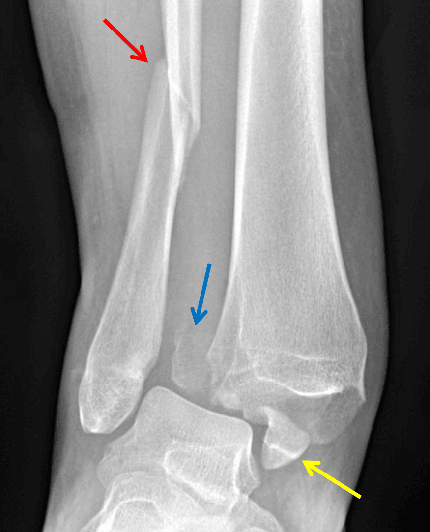
Weber type C fracture - Angulated distal fibular fracture (red arrow) with associated tibiofibular syndesmotic injury and displaced posterior (blue arrow) and medial (yellow arrow) malleolar fractures.
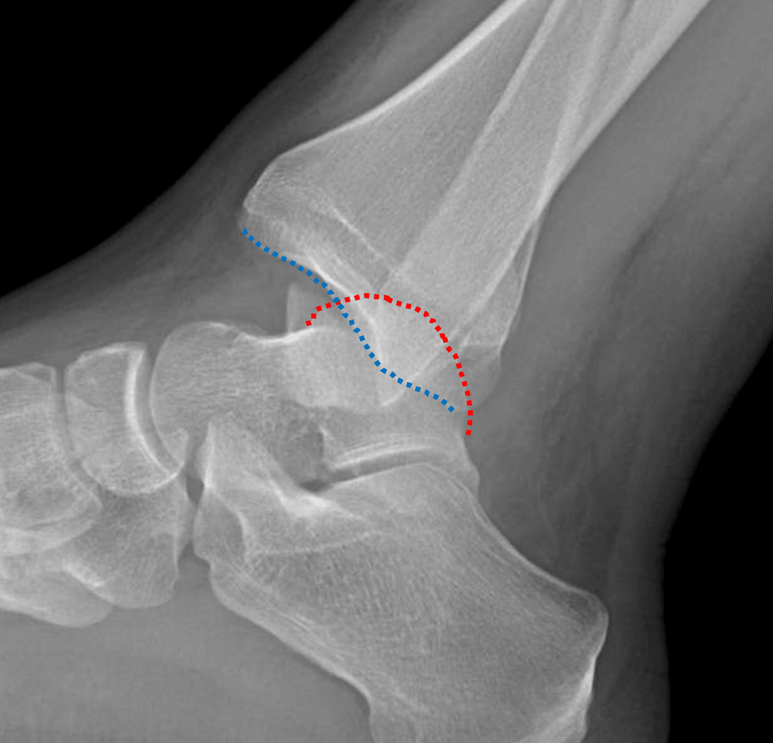
Posterior dislocation of the ankle with the talar dome (red dotted line) positioned posterior to the tibial plafond (blue dotted line).
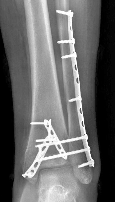
Radiograph in this patient after ORIF.