Use mouse wheel, arrow keys or left click (with scroll tool selected) to scroll
ui.case.use_touch_gestures
DICOM HelpSource: Local (us-east1-c)
Keyboard shortcuts (Alt+K)
Demographics:
5 days old, Male
Indication:
Rigid abdomen (born at 29 weeks)
Findings
- Large volume pneumoperitoneum
- Featureless nondistended loops of bowel are seen throughout the abdomen
- Diffuse granular airspace opacities at the lung bases
- Umbilical venous catheter tip projects over the right atrium
- Umbilical arterial catheter tip projects at the level of T5-T6
Diagnosis
Pneumoperitoneum
Sample Report
Large volume pneumoperitoneum.
Featureless nondistended loops of bowel are seen throughout the abdomen without convincing pneumatosis.
Diffuse granular airspace opacities at the lung bases, which may relate to respiratory distress syndrome.
Umbilical venous catheter tip projects over the right atrium.
Umbilical arterial catheter tip projects at the level of T5-T6.
Discussion
- Bowel perforation in neonates most commonly occurs in the distal ileum or proximal colon
- Lateral decubitus views increase the sensitivity for free air with left side down preferred so that free air will layer along the smooth contour of the liver, making it easier to detect
- Other radiographic findings of pneumoperitoneum include:
- Football sign - generalized hyperlucency of the abdomen with the falciform ligament resembling the lacing of a football
- Rigler sign - gas along bowel walls makes both the inner and outer margins of the wall visible
- Continuous appearance of the diaphragm (remember that pneumomediastinum can also do this)
Annotated Images & Illustrations
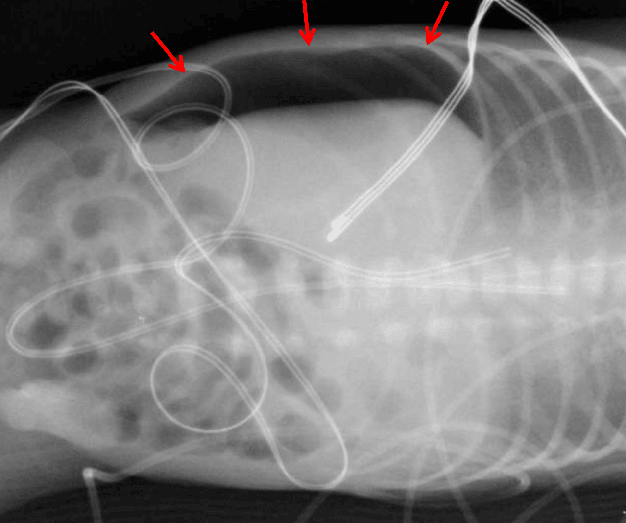
Red arrows: large volume pneumoperitoneum on left side down lateral decubitus view.