Keyboard shortcuts (Alt+K)
Demographics:
7 days old, Female
Indication:
Melena (term delivery)
Findings
- Extensive portal venous gas
- Mottled and curvilinear lucencies along loops of bowel in the left upper quadrant
- No definite evidence of pneumoperitoneum
- Nonfocal gaseous distension of bowel loops throughout the abdomen
- Enteric tube tip overlies the stomach
Diagnosis
Necrotizing enterocolitis (NEC)
Sample Report
Findings concerning for necrotizing enterocolitis with extensive portal venous gas as well as mottled and curvilinear lucencies along loops of bowel in the left upper quadrant suspicious for pneumatosis.
No definite evidence of pneumoperitoneum. However, recommend left side down decubitus view for a more sensitive evaluation.
Nonfocal gaseous distension of bowel loops throughout the abdomen.
Enteric tube tip overlies the stomach.
Discussion
- Look for the following imaging findings to raise suspicion for necrotizing enterocolitis:
- Bowel gas pattern: often mildly dilated and nonfocal. If bowel gas pattern remains unchanged between radiographs, this is concerning for bowel dysfunction and even developing ischemia
- Pneumatosis: look for rounded or linear lucencies centered within the bowel wall. When extensive, bubbly lucencies can look similar to mottled lucencies in stool, so determining whether the lucencies are intraluminal (benign) or intramural (bad) is very important
- Portal venous gas: while this can occur anywhere within the portal venous tree, it is most easily recognized within the liver on radiographs. Look for branching lucencies extending to the periphery of the liver (unlike pneumobilia which is typically only seen near the hilum)
- Pneumoperitoneum: make sure to get a lateral decubitus view or even a cross-table lateral view with the patient supine if necessary to increase your sensitivity for detecting free air
- However, be aware that none of these findings are necessary for the diagnosis, nor do they perfectly correlate with disease severity
Annotated Images & Illustrations
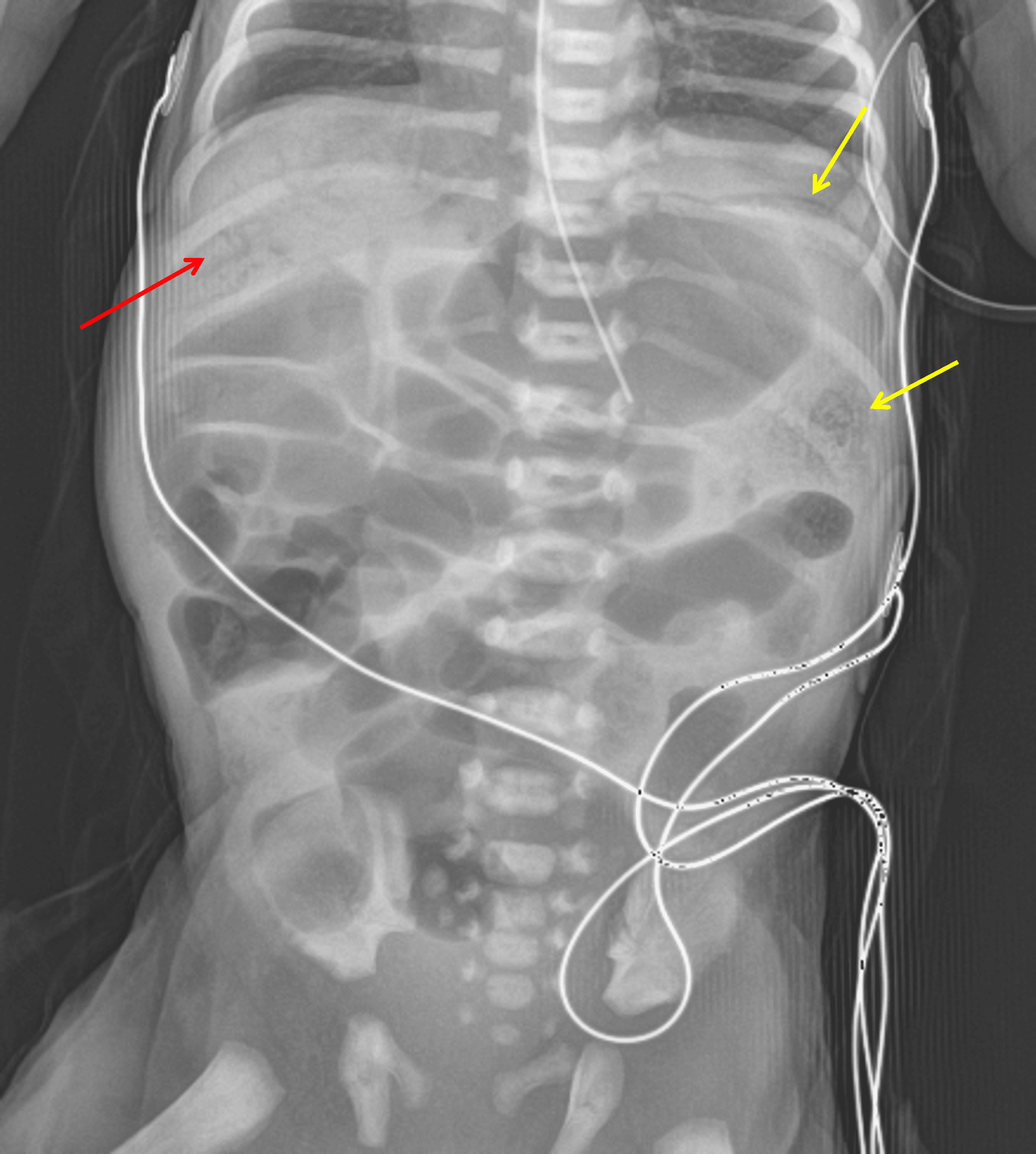
Red arrow: multiple branching lucencies overlying the liver consistent with portal venous gas. Yellow arrows: curvilinear and mottled lucencies along bowel loops in the left upper quadrant.