Use mouse wheel, arrow keys or left click (with scroll tool selected) to scroll
ui.case.use_touch_gestures
DICOM HelpSource: Local (us-east1-c)
Findings
- Occlusive/near occlusive dural venous sinus thrombosis extending from the distal superior sagittal sinus into the left transverse sinus, left sigmoid sinus, left jugular bulb, and proximal left internal jugular vein. Thrombus also slightly extends into the proximal right transverse sinus and involves multiple left temporal cortical veins including the vein of Labbé
- Small area of cortical and subcortical T2/FLAIR hyperintensity in the left middle temporal gyrus with faint cortical restricted diffusion
- No acute hemorrhage
- No significant mass effect or hydrocephalus
- No evidence of proximal intracranial arterial occlusion
Diagnosis
Venous sinus thrombosis
Sample Report
Occlusive/near occlusive dural venous sinus thrombosis extending from the distal superior sagittal sinus into the left transverse sinus, left sigmoid sinus, left jugular bulb, and proximal left internal jugular vein. Thrombus also slightly extends into the proximal right transverse sinus and involves multiple left temporal cortical veins including the vein of Labbé.
Associated small area of congestive edema in the left middle temporal gyrus with faint cortical restricted diffusion possibly representing early ischemic changes.
No acute hemorrhage.
No significant mass effect or hydrocephalus.
No evidence of proximal intracranial arterial occlusion.
Discussion
- Think about sinus thrombosis when you see patterns of ischemia atypical for thrombotic infarcts, including:
- Superior sagittal sinus thrombosis -> symmetric parasagittal regions
- Vein of Labbe -> temporal lobe
- Deep venous thrombosis (straight sinus, internal cerebral veins) -> bilateral thalami
- Venous thrombosis progresses to ischemia much slower than arterial thrombosis, generally somewhere on the range of several days to several weeks. If the thrombus is treated/removed, the area of related edema will often resolve without progressing to infarct
- Deep venous infarcts are more serious than dural venous infarcts, particularly due to potential resulting thalamic ischemia
- Just like arterial infarcts, be sure to mention any associated mass effect or hemorrhagic transformation
Annotated Images & Illustrations
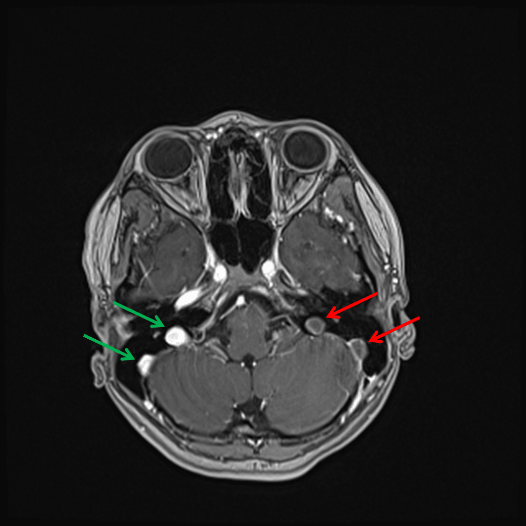
Nonopacification of the left sigmoid sinus and left jugular bulb (red arrows) in contrast to the normally opacified structures on the right (green arrows) on this postcontrast image.
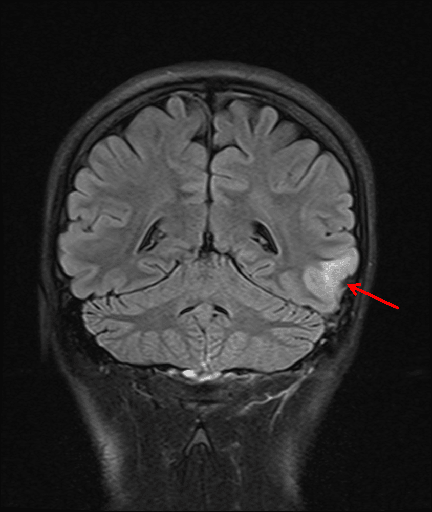
Small area of FLAIR signal hyperintensity in the left middle temporal gyrus (red arrow).
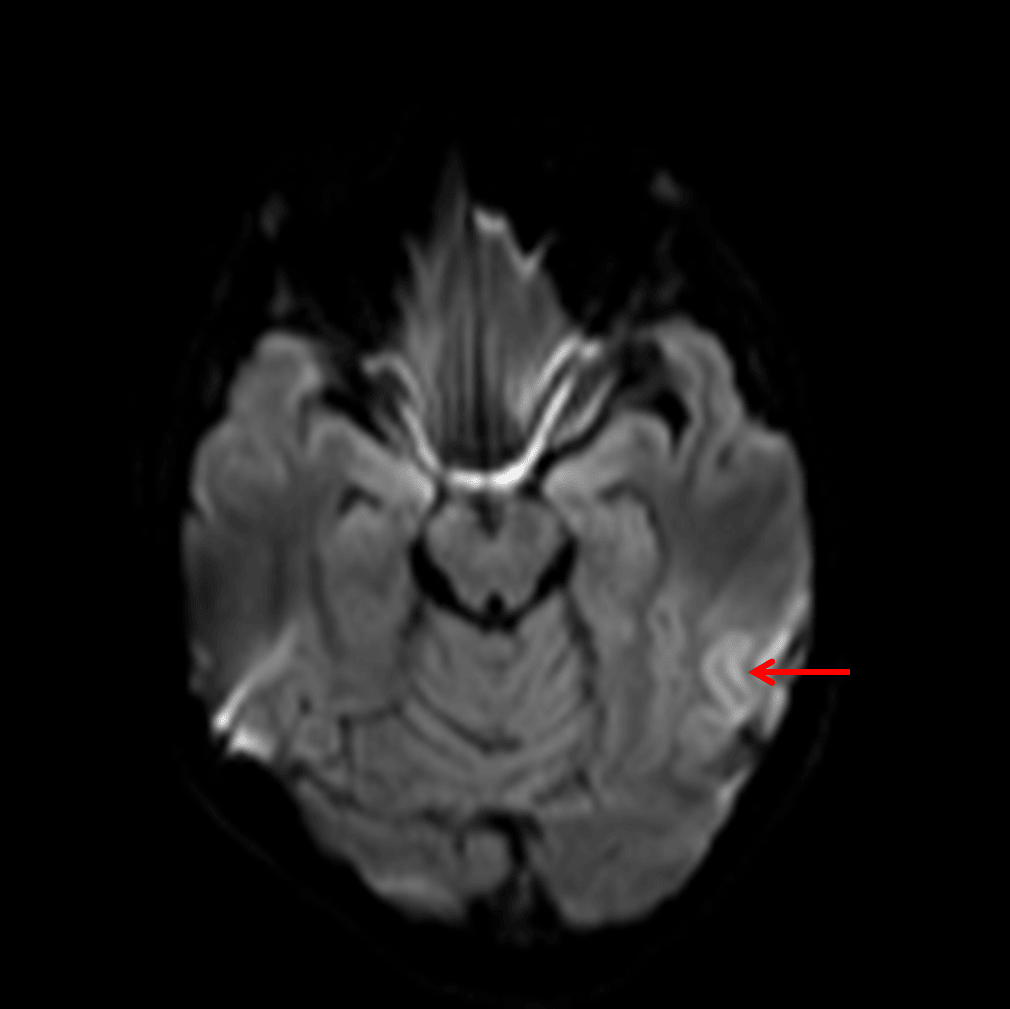
Faint cortical diffusion signal hyperintensity (red arrow).
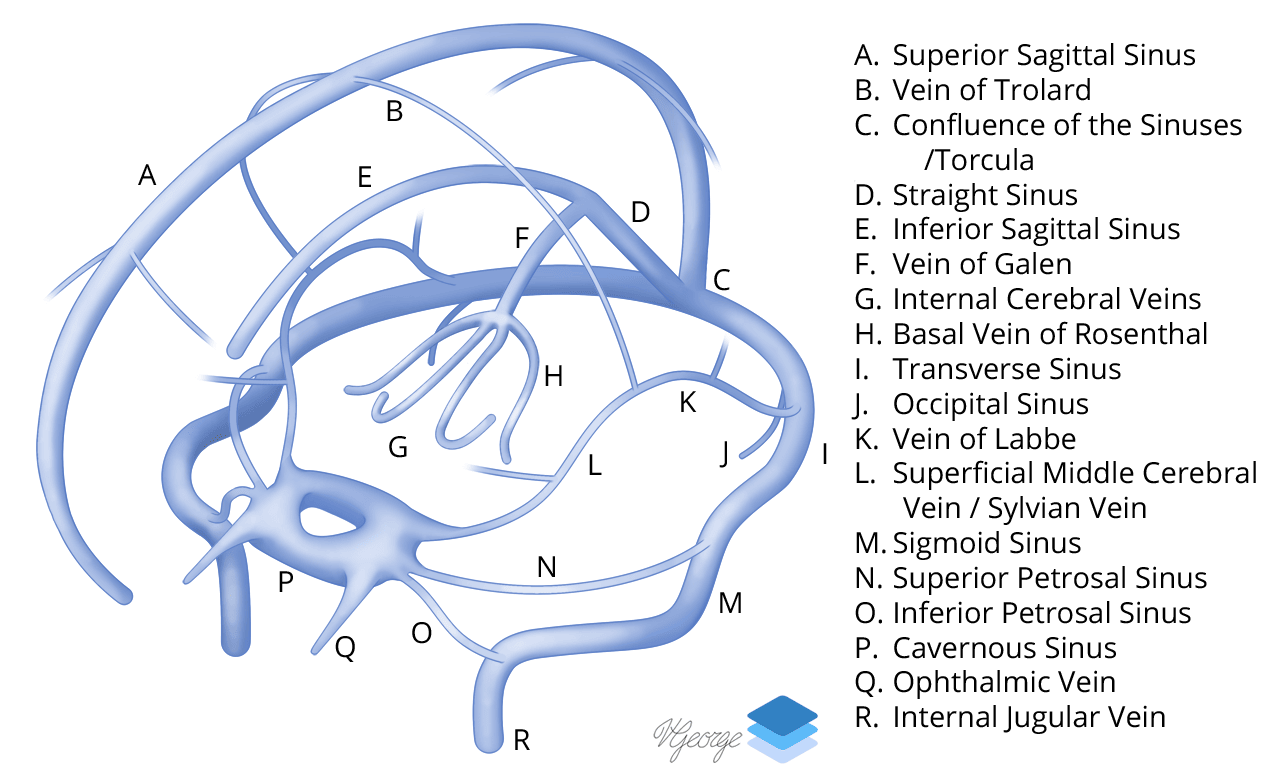
Venous Sinuses.
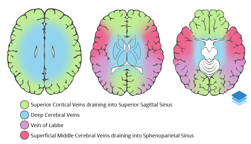
Approximations of venous vascular territories of the brain.