Use mouse wheel, arrow keys or left click (with scroll tool selected) to scroll
ui.case.use_touch_gestures
DICOM HelpSource: Local (us-east1-c)
Keyboard shortcuts (Alt+K)
Demographics:
34 years old, Male
Indication:
Shoulder injury
Findings
- Posterior shoulder dislocation
- Acute reverse Hill-Sachs impaction fracture of the anterior humeral head
- Acute minimally displaced reverse Bankart fracture of the posterior glenoid
Diagnosis
Posterior shoulder dislocation with reverse Hill-Sachs and Bankart fractures
Sample Report
Posterior shoulder dislocation with an acute reverse Hill-Sachs impaction fracture and an acute minimally displaced reverse Bankart lesion.
Acromioclavicular joint alignment is maintained.
Discussion
- Shoulder dislocations come in three major varieties: anterior, posterior, and inferior, with anterior being by far the most common. Here are some radiographic clues for each type of dislocation:
- Anterior: humeral head inferior and medial to the glenoid
- Posterior: humeral head may appear correctly positioned relative to the glenoid on AP view. Shoulder will classically be fixed in internal rotation (the so-called “lightbulb” sign)
- Inferior: humeral head inferior to the glenoid with the arm often fixed in abduction (raised over the head, aka "luxatio erecta")
- Associated fractures:
- Hill-Sachs
- Occurs along the posterolateral humeral head where the humeral head impacts on the glenoid
- Best seen on internal rotation views
- Reverse Hill-Sachs from posterior dislocation -> impaction fracture of the inferomedial humeral head, which results in a “trough” sign on radiographs
- Bankart
- Anterior rim of the glenoid, often difficult to see on radiographs
- Reverse Bankart (posterior rim) with posterior shoulder dislocation
- Hill-Sachs
Annotated Images & Illustrations
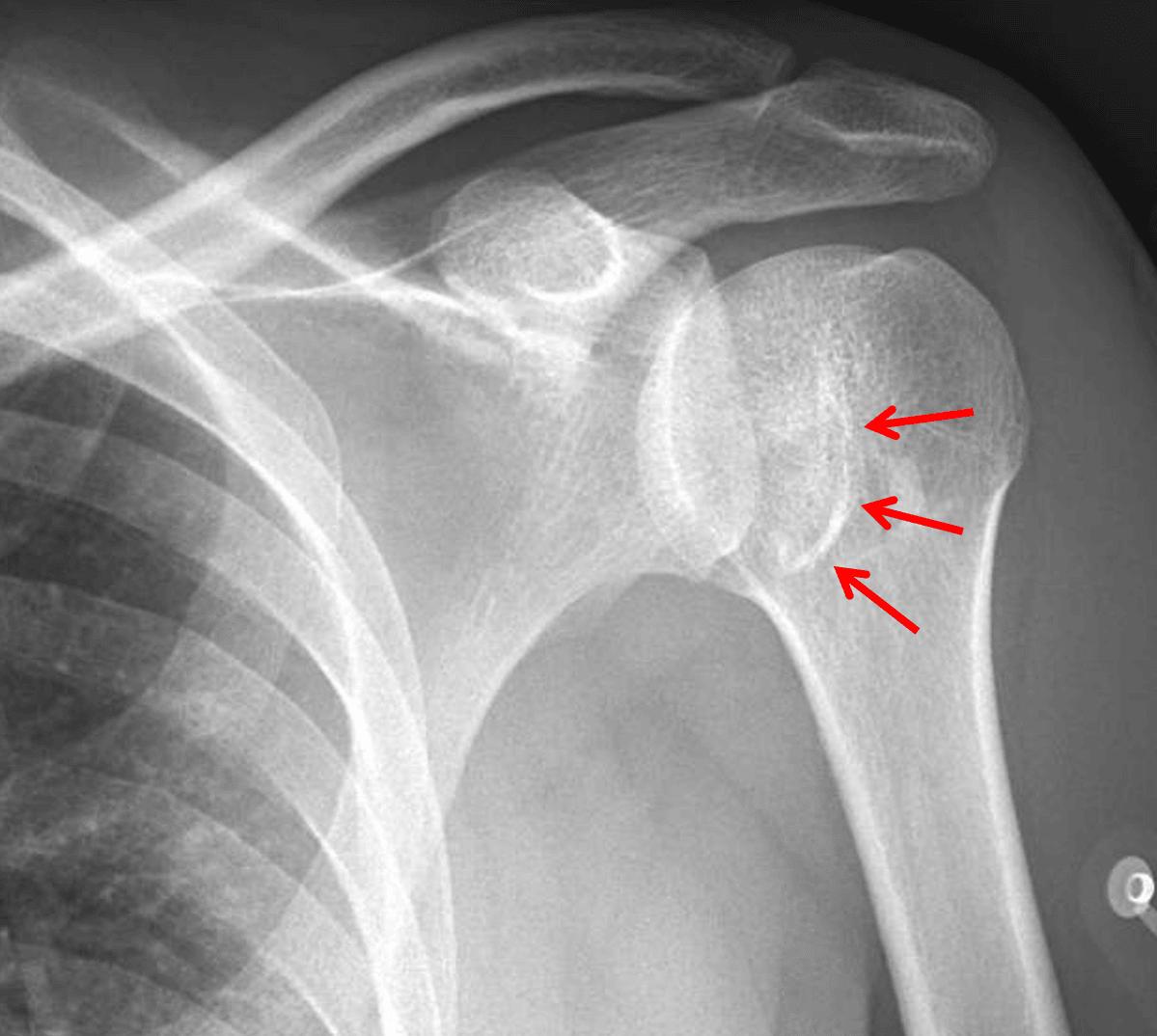
Red arrows: reverse Hill-Sachs fracture of the inferomedial humeral head resulting in a trough sign on frontal views.
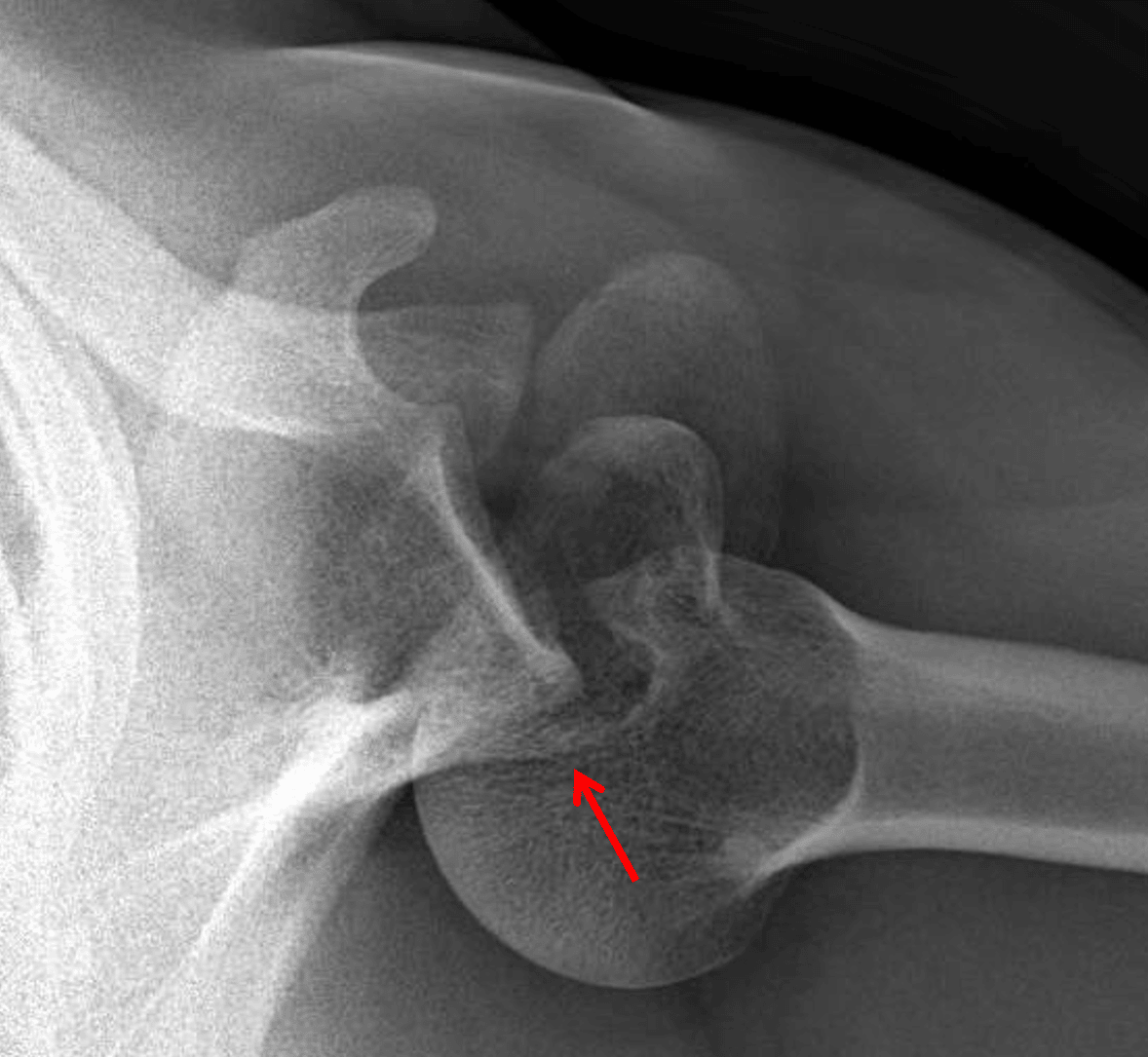
Red arrow: posterior shoulder dislocation with minimally displaced fracture of the posterior glenoid, best shown on this axillary view.