Use mouse wheel, arrow keys or left click (with scroll tool selected) to scroll
ui.case.use_touch_gestures
DICOM HelpSource: Local (us-east1-c)
Keyboard shortcuts (Alt+K)
Demographics:
55 years old, Male
Indication:
Chest pain
Findings
- Mass-like opacity along the left upper mediastinum, which is superimposed over the left pulmonary artery on the PA projection and overlaps the great vessels on the lateral projection
- No evidence of acute cardiopulmonary disease
Diagnosis
Middle mediastinal mass (duplication cyst)
Sample Report
Left middle mediastinal mass. Recommend CT for further evaluation.
Otherwise no evidence of acute cardiopulmonary disease.
Discussion
- It is important to try to separate mediastinal masses based on location in the anterior, middle, or posterior mediastinum
- A recent publication by the International Thymic Malignancy Interest Group (ITMIG) defines the three mediastinal compartments as prevascular (bounded posteriorly by the pericardium), visceral (bounded anteriorly by the pericardium and posteriorly by an imaginary line drawn through each thoracic vertebral body 1 cm from its anterior margin), and paravertebral
- Think about the following categories with middle (visceral) mediastinal masses:
- vascular pathology - e.g. dissection, aneurysm, variant anatomy
- lymph nodes
- tumors - especially cardiac or pericardial
- incidental - pericardial cysts, duplication cysts
- esophageal pathology: tumor, dilation, hiatal hernia
- This case turned out to be a benign duplication cyst (see CT below)
Annotated Images & Illustrations
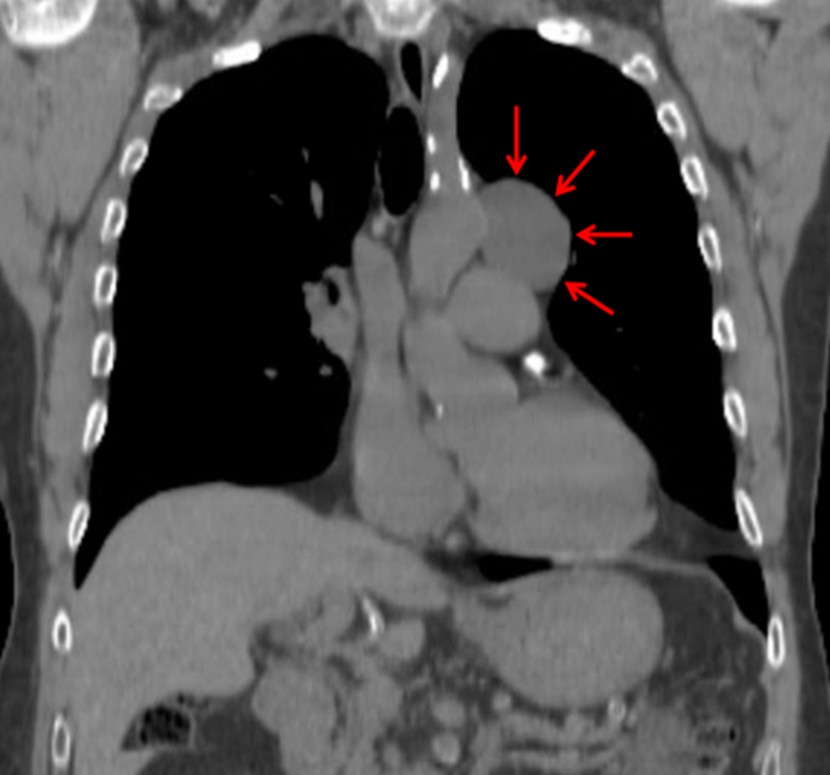
CT demonstrated a middle mediastinal mass most consistent with a benign duplication cyst (red arrows).