Use mouse wheel, arrow keys or left click (with scroll tool selected) to scroll
ui.case.use_touch_gestures
DICOM HelpSource: Local (us-east1-c)
Keyboard shortcuts (Alt+K)
Demographics:
44 years old, Female
Indication:
Myasthenia Gravis
Findings
- Smoothly marginated soft tissue mass adjacent to the right heart border, likely in the anterior mediastinum
- No evidence of acute cardiopulmonary disease
Diagnosis
Anterior mediastinal mass (thymoma)
Sample Report
No evidence of acute cardiopulmonary disease.
Smoothly marginated soft tissue mass adjacent to the right heart border, likely in the anterior mediastinum. Recommend obtaining a chest CT for further evaluation.
Discussion
- CT was obtained and showed an anterior mediastinal mass that turned out to be a thymoma
- The boundaries of the anterior mediastinum include the right and left heart borders and ascending aorta. Therefore, bulging of these contours can be seen with an anterior mediastinal mass
- Consider the following when you see an anterior (prevascular) mediastinal mass:
- Thymic pathology - thymoma, carcinoma, cyst
- Enlarged lymph nodes - e.g. lymphoma
- Germ cell tumor
- Thyroid enlargement/mass
Annotated Images & Illustrations
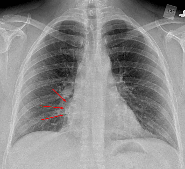
Approximately 4 cm mass in the mediastinum (red arrows) adjacent to the right heart border.
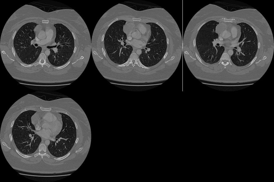
Subsequent CT shows an anterior mediastinal mass most consistent with a thymoma.
References