Use mouse wheel, arrow keys or left click (with scroll tool selected) to scroll
ui.case.use_touch_gestures
DICOM HelpSource: Local (us-east1-c)
Keyboard shortcuts (Alt+K)
Demographics:
79 years old, Female
Indication:
Shortness of breath
Findings
- Triangular opacity at the posteromedial right lung base with loss of the silhouette of the medial and posterior aspect of the right hemidiaphragm
- Evidence of volume loss in the right hemithorax including elevation of the right hemidiaphragm, inferior positioning of the right hilum, rightward mediastinal shift, and crowding of many right ribs
- Possible right hilar fullness
- Likely chronic interstitial coarsening
- Left upper extremity PICC tip projects over the superior cavoatrial junction
- Feeding tube courses beyond the inferior border of the image
- Anterior wedging of a midthoracic vertebral body with associated focal kyphosis
- Thoracic levoscoliosis
- Osteopenia
Diagnosis
Right lower lobe collapse
Sample Report
Right lower lobe collapse with question of right hilar fullness. Recommend chest CT to exclude an obstructing hilar mass. Superimposed aspiration/pneumonia at the right lung base is not excluded, though the right basilar opacification could be entirely explained by lobar collapse.
Discussion
- The right lower lobe usually collapses to the inferior/posterior/medial aspect of the right hemithorax, resulting in loss of the medial and posterior silhouette of the right hemidiaphragm. The right heart border silhouette is preserved, which helps distinguish right lower lobe collapse from right middle lobe collapse
Annotated Images & Illustrations
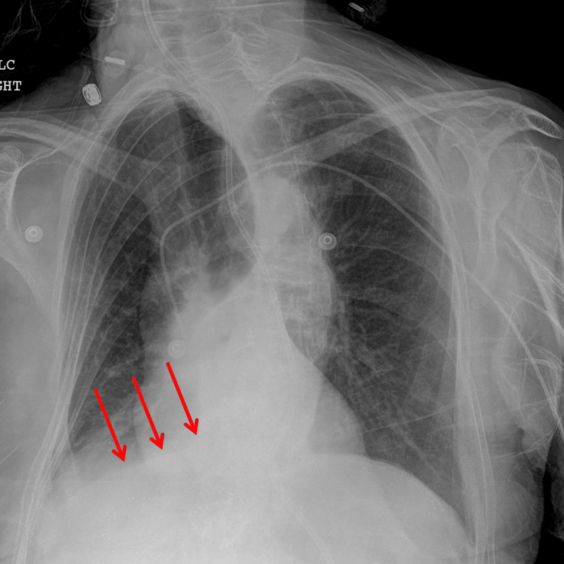
Red arrows: right lower lobe collapse.
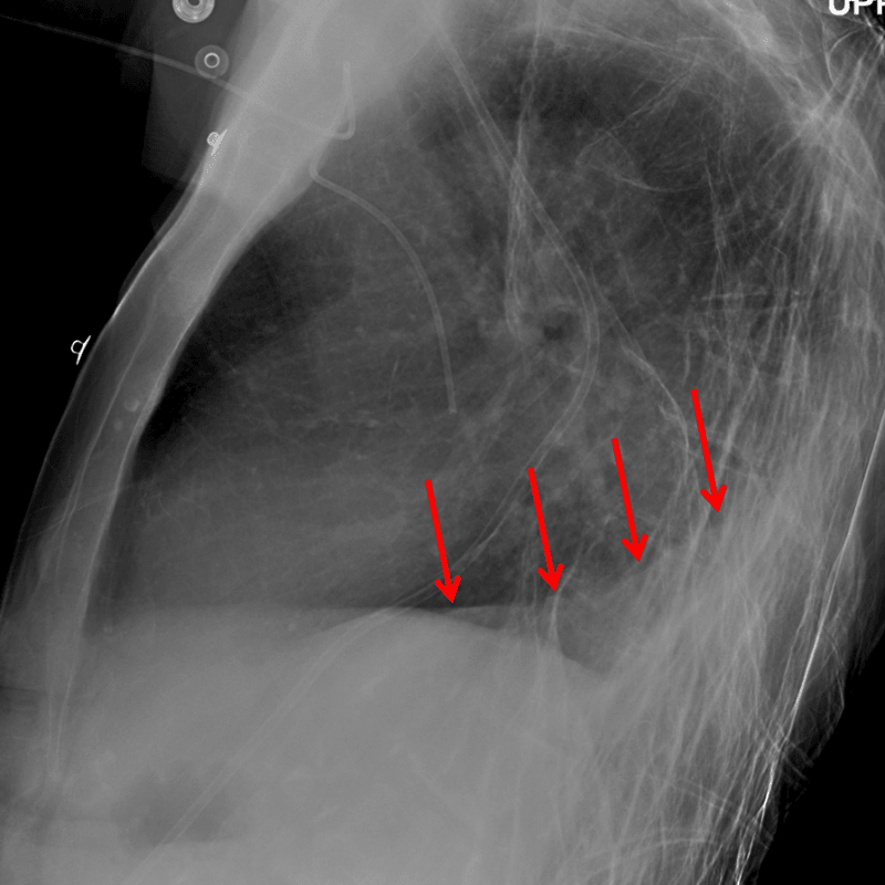
Red arrows: right lower lobe collapse.
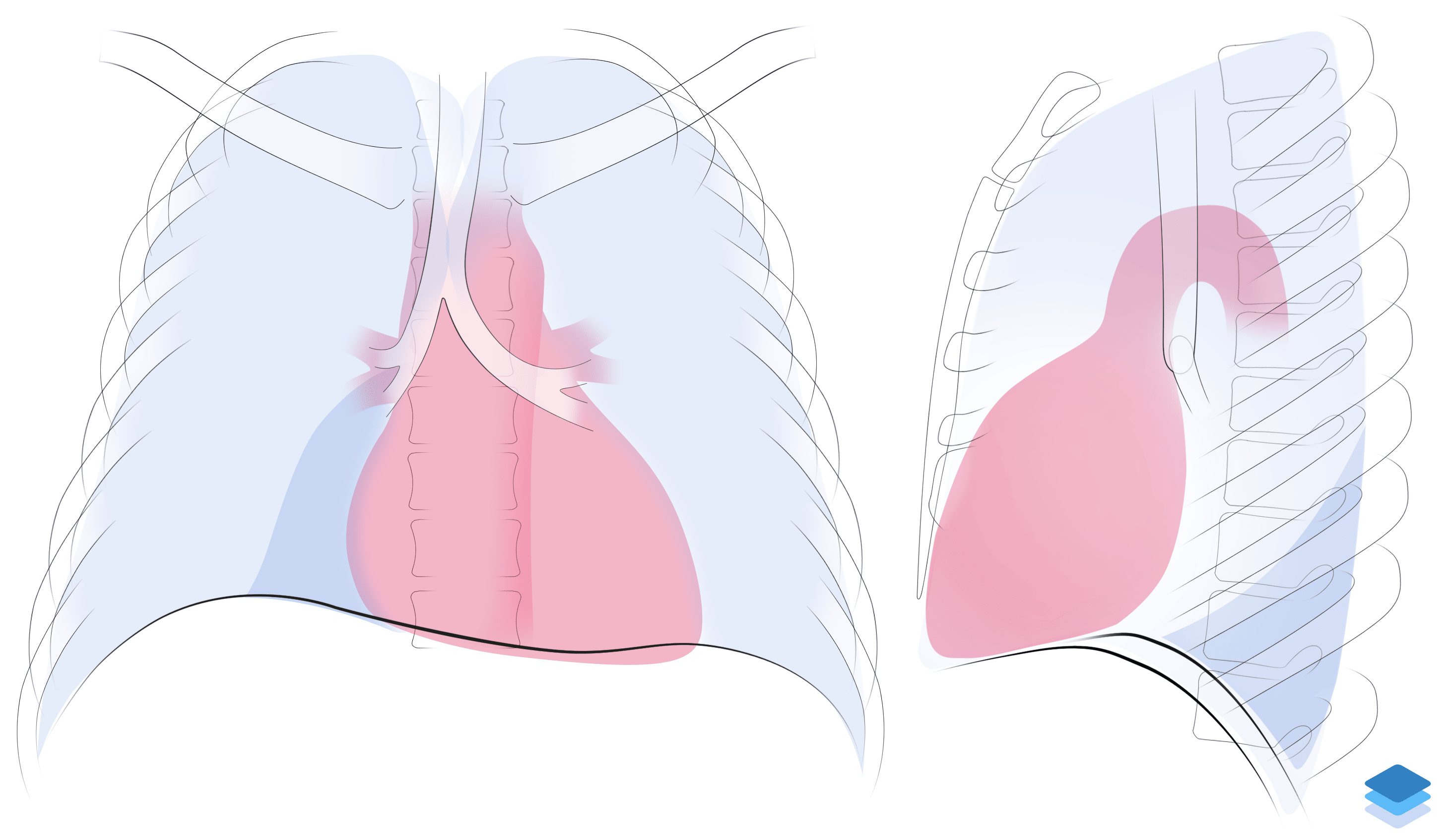
Right lower lobe collapse. Illustration by Valerie George, MD