Use mouse wheel, arrow keys or left click (with scroll tool selected) to scroll
ui.case.use_touch_gestures
DICOM HelpSource: Local (us-east1-c)
Keyboard shortcuts (Alt+K)
Demographics:
25 years old, Male
Indication:
Right-sided chest pain, cough
Findings
Chest radiograph
- Hazy opacification in the right middle lobe which likely represents a combination of atelectasis (given mild tenting of the right hemidiaphragm) and airspace filling
CT
Chest
- Mild volume loss in the right middle lobe with groundglass opacification throughout the lobe and consolidation in the lateral segment
- Additional scattered groundglass opacities in the right lower lobe and lingula
- Mild dependent right lower lobe atelectasis
- Small right pleural effusion
- Multiple mildly enlarged mediastinal lymph nodes, likely reactive
Upper abdomen
- No acute findings
MSK
- No acute findings
Diagnosis
Lobar pneumonia
Sample Report
Right middle lobe pneumonia.
Additional scattered groundglass opacities in the right lower lobe and lingula are likely also infectious in etiology.
Small right pleural effusion.
Discussion
- Lobar pneumonia (typical pneumonia) typically manifests as a mixture of groundglass opacification and consolidation as infection spreads between contiguous alveoli via pores of Kohn and canals of Lambert
- The diagnosis of pneumonia is clinical, so the primary role of the radiologist is to look for complications:
- If a pleural effusion is present, make sure to look for pleural thickening, loculation, or pleural gas to suggest the presence of an empyema
- Severe infections can compromise the vascular supply, leading to tissue necrosis and cavitation - make sure to look for intraparenchymal collections/abscess
- This case has coexisting pneumonia and atelectasis in the right middle lobe, which accounts for the confusing volume loss not typically seen with infection alone
Annotated Images & Illustrations
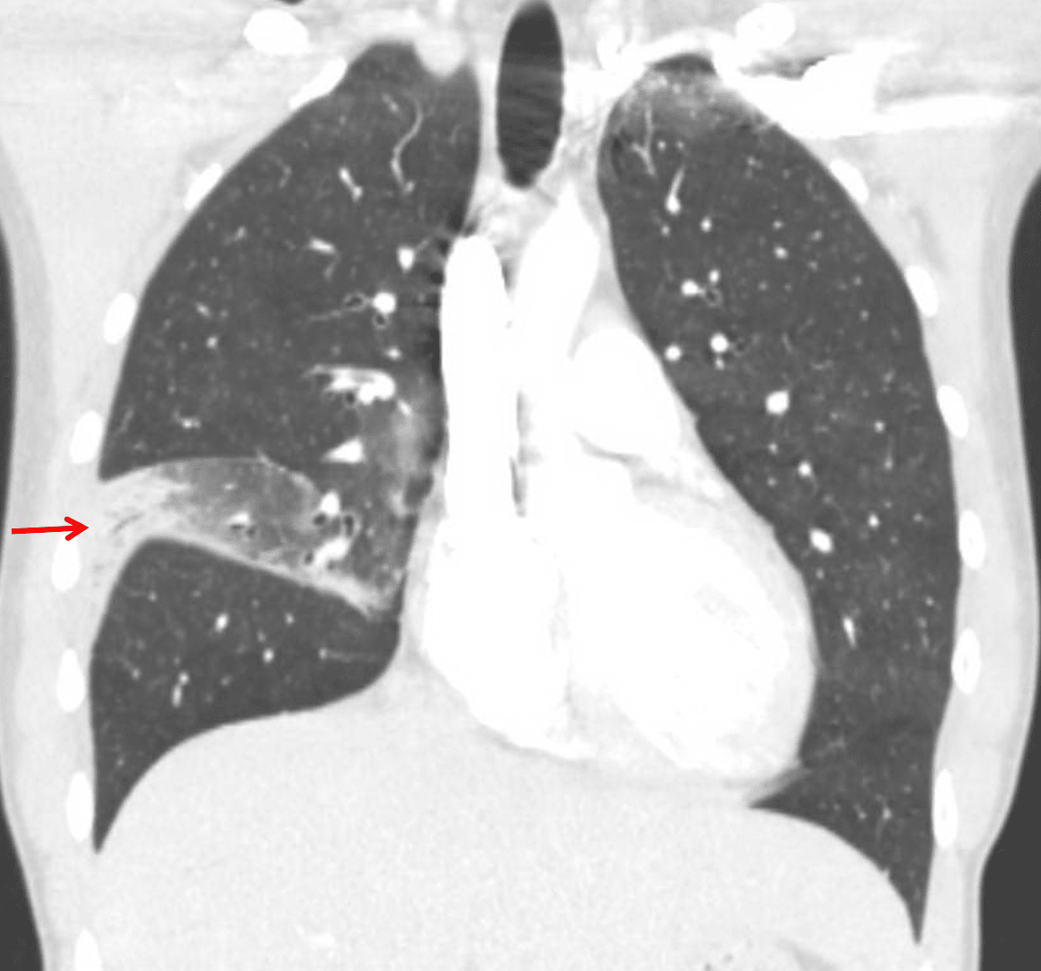
Lobar pneumonia: consolidation in the lateral segment of the right middle lobe with associated volume loss in the right middle lobe.