患者信息:
16 岁, 男性
检查指征:
踝关节损伤伴明显畸形
诊断
Weber C型骨折
踝关节脱位
示例报告
急性斜行腓骨远端骨折,短缩约1 cm伴后外侧成角(Weber C型),伴远端胫腓联合增宽及距胫关节后方脱位。MRI可进一步评估韧带损伤范围。
急性分离性内踝骨折,外侧移位约2 cm。
急性轻度粉碎性后踝骨折,外侧移位约2 cm。
讨论
- 虽然存在更复杂的踝关节骨折分型系统,但至少应熟悉Weber分型:
- Weber A型:外踝骨折位于距骨穹窿水平以下——通常稳定,保守治疗
- Weber B型:外踝骨折线于距骨穹窿水平内侧出口——如内侧结构完整则稳定:注意寻找内侧间隙增宽(提示三角韧带损伤)和远端胫腓联合增宽
- Weber C型:外踝骨折位于距骨穹窿水平以上——通常不稳定,因为这些骨折伴有远端胫腓联合损伤
- 如同桡骨和尺骨一样,将胫骨和腓骨视为环形结构。如果在一处看到损伤(骨折或关节半脱位),需检查环形结构的其余部分是否有额外损伤。如果仅看到踝穴内侧间隙增宽,确保获取胫腓骨全长片以寻找腓骨近端(Maisonneuve)骨折
带注释的图像和插图
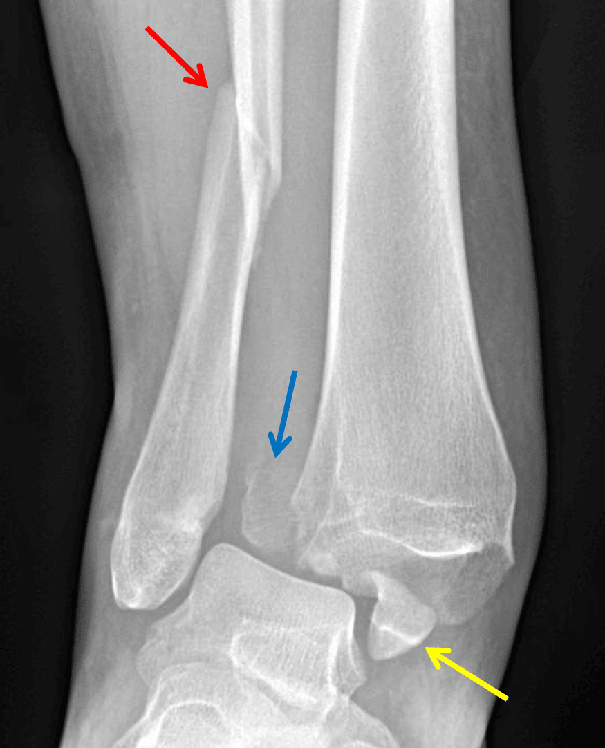
Weber type C fracture - Angulated distal fibular fracture (red arrow) with associated tibiofibular syndesmotic injury and displaced posterior (blue arrow) and medial (yellow arrow) malleolar fractures.
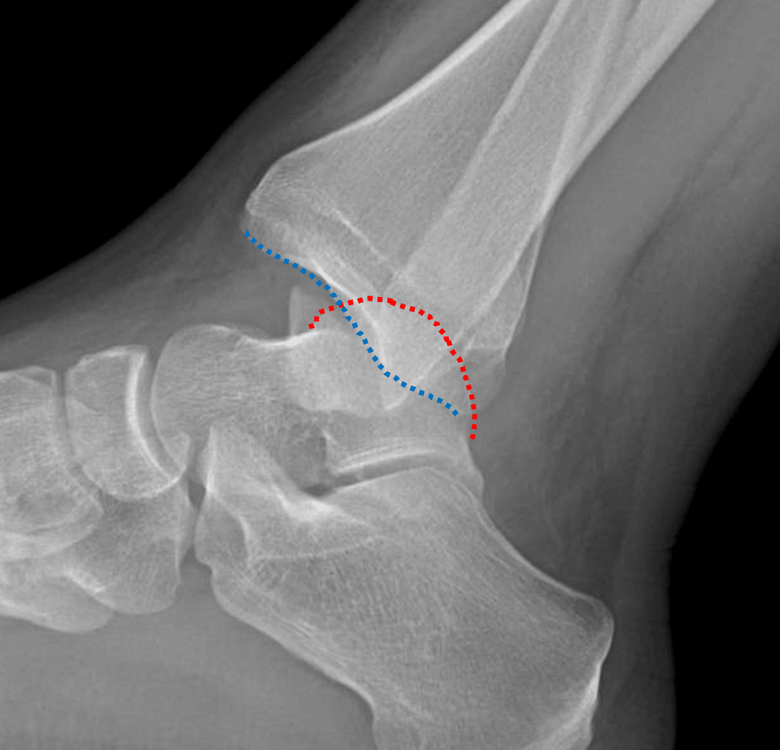
Posterior dislocation of the ankle with the talar dome (red dotted line) positioned posterior to the tibial plafond (blue dotted line).
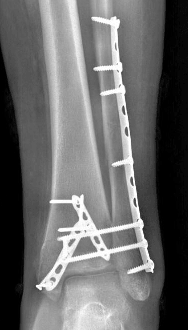
Radiograph in this patient after ORIF.