1/22
患者信息:
13 岁, 女性
检查指征:
癫痫发作
影像所见
诊断
静脉窦血栓形成
示例报告
闭塞性/近闭塞性硬脑膜静脉窦血栓形成,从上矢状窦远端延伸至左侧横窦、左侧乙状窦、左侧颈静脉球及左侧颈内静脉近端。血栓亦轻度延伸至右侧横窦近端并累及多条左侧颞叶皮层静脉,包括Labbé静脉。
左侧颞中回相关小面积充血性水肿,伴微弱皮层弥散受限,可能代表早期缺血改变。
无急性出血。
无显著占位效应或脑积水。
无近端颅内动脉闭塞征象。
讨论
- 当看到不典型于血栓性梗死的缺血模式时,应考虑静脉窦血栓形成,包括:
- 上矢状窦血栓 -> 双侧旁矢状区
- Labbé静脉 -> 颞叶
- 深静脉血栓(直窦、大脑内静脉)-> 双侧丘脑
- 静脉血栓导致缺血的进展远慢于动脉血栓,通常在数天至数周范围内。如果血栓被治疗/清除,相关水肿区域通常会消退而不进展为梗死
- 深静脉梗死比硬脑膜静脉梗死更为严重,尤其是由于可能导致丘脑缺血
- 与动脉性梗死一样,务必提及任何相关的占位效应或出血性转化
带注释的图像和插图
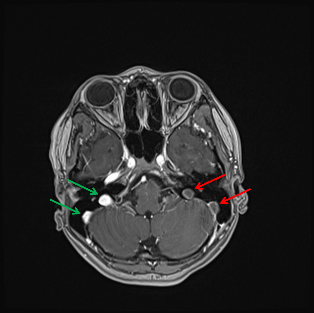
Nonopacification of the left sigmoid sinus and left jugular bulb (red arrows) in contrast to the normally opacified structures on the right (green arrows) on this postcontrast image.
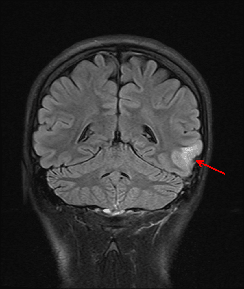
Small area of FLAIR signal hyperintensity in the left middle temporal gyrus (red arrow).
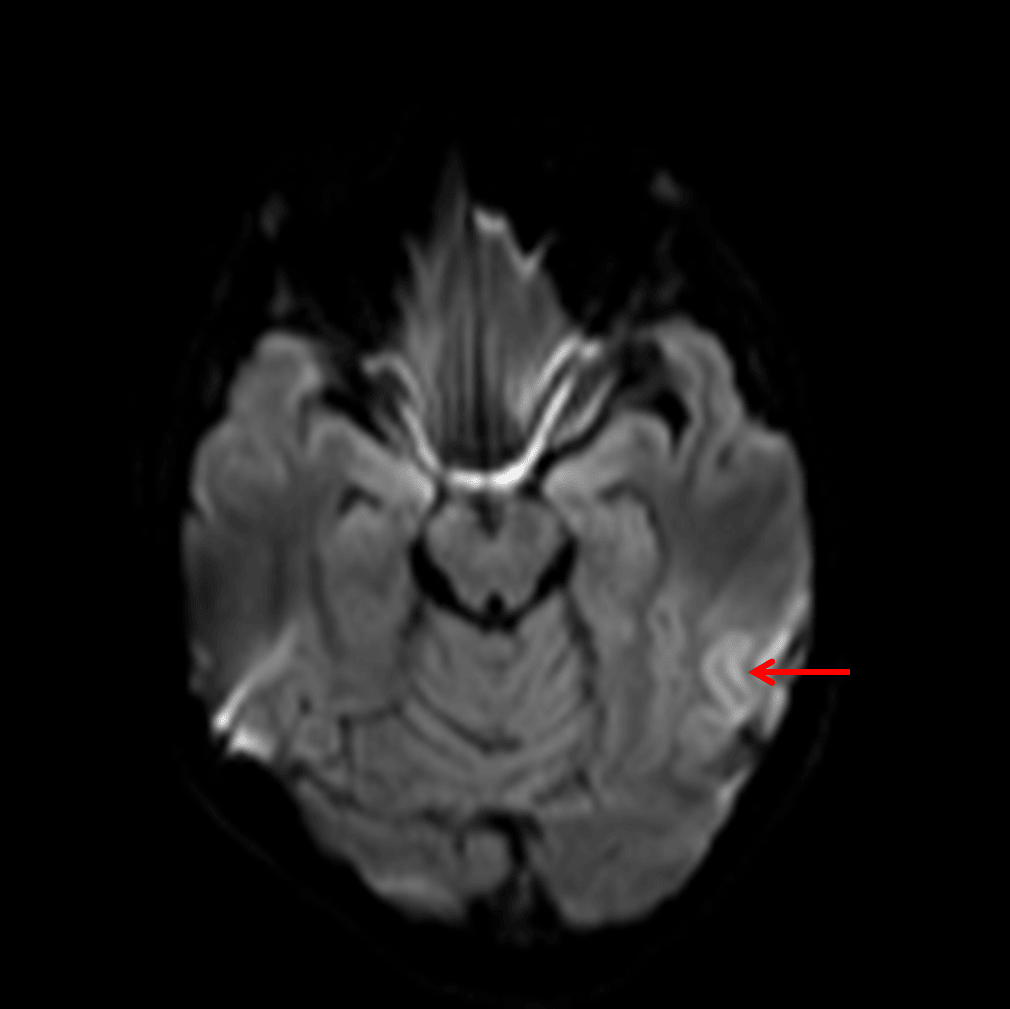
Faint cortical diffusion signal hyperintensity (red arrow).
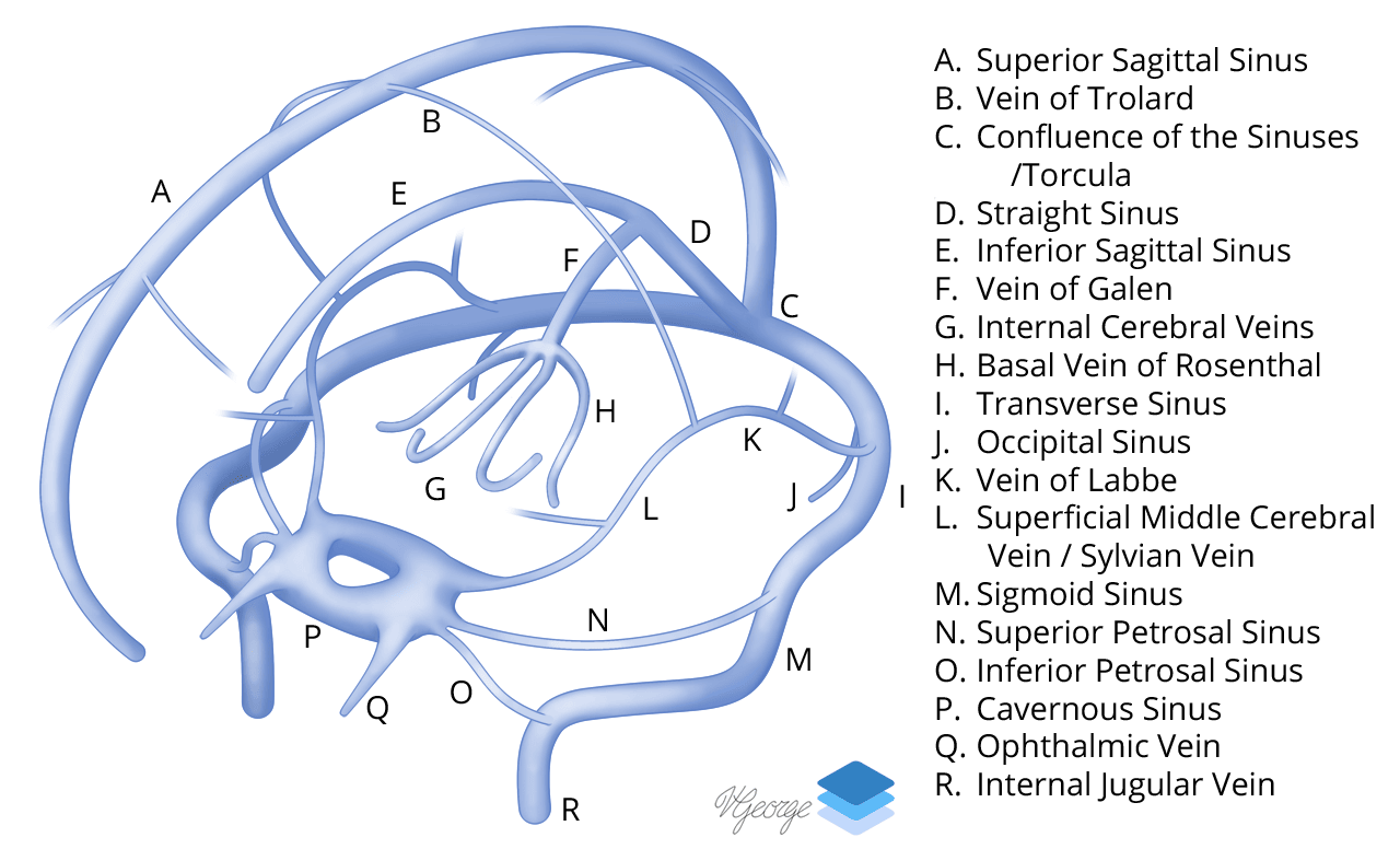
Venous Sinuses.
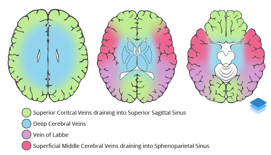
Approximations of venous vascular territories of the brain.