Demographics:
9 years old, Male
Indication:
Precocious puberty
Findings
- Suprasellar mass measuring 3.5 x 2 x 3.3 cm with heterogeneous internal T1 and T2 signal
- Nodular enhancing component superiorly measuring up to 1.8 cm
- The optic chiasm appears to be incorporated within the mass
- The pituitary gland is visualized separate from the mass
- The mass contacts the right A1 ACA segment, both supraclinoid internal carotid arteries, the basilar artery, the posterior communicating arteries, and the P1 segments of both PCAs
- The mass also contacts and deforms the floor of the third ventricle
Annotated Images & Illustrations
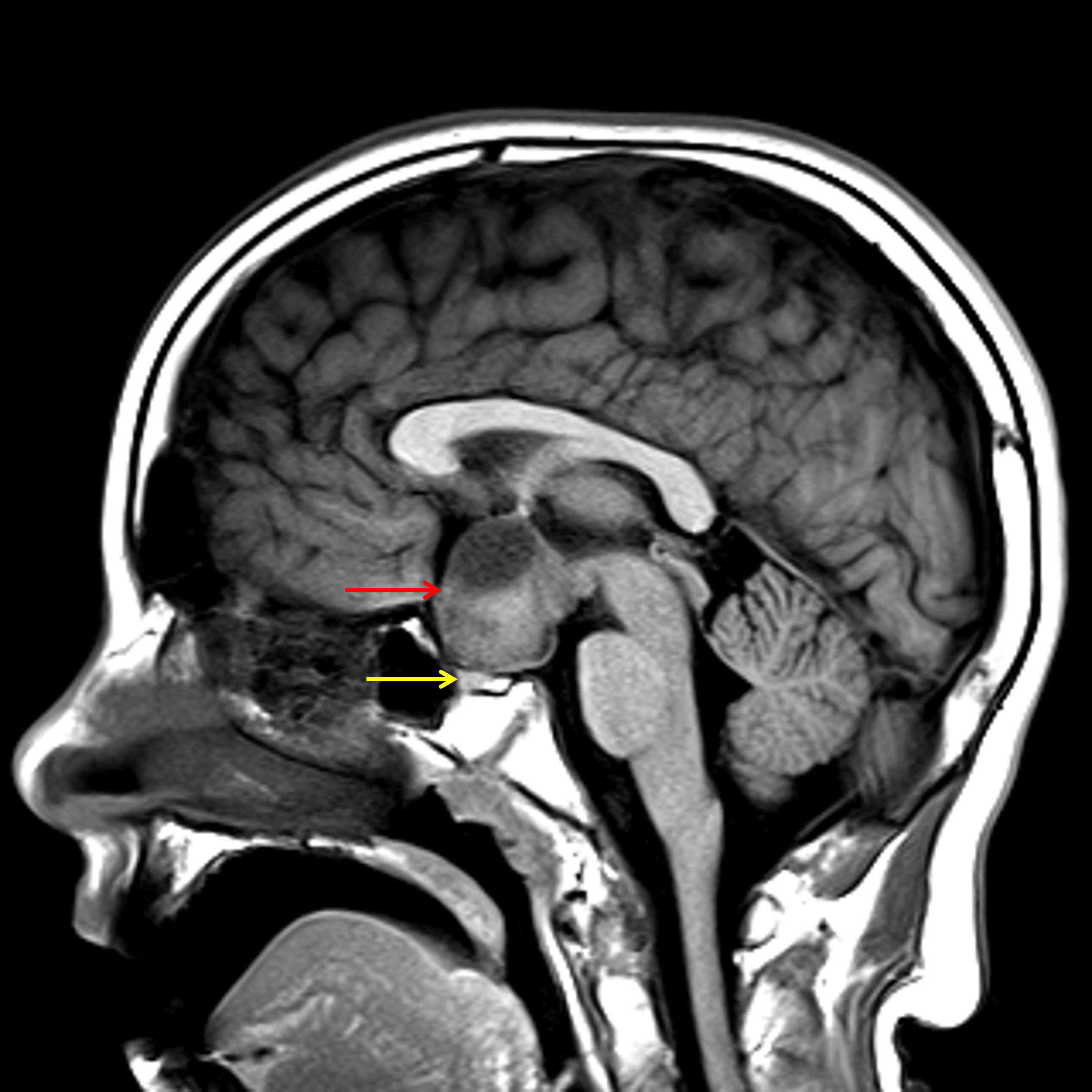
Heterogeneous suprasellar mass (red arrow) which is seen separate from the pituitary gland (yellow arrow).
Diagnosis
Optic pathway glioma (pilocytic astrocytoma)
Differential Diagnosis
Become a PRO member to unlock the differential diagnosis
Pearls
Become a PRO member to unlock the pearls