Demographics:
39 years old, Female
Indication:
Seizure
Findings
- Peripherally-located mass in the anterior aspect of the left frontal lobe measuring 4.5 x 5.5 x 5 cm
- The mass is largely nonenhancing, though there may be minimal nodular enhancement along its anteromedial margin
- Central cystic component
- No corresponding restricted diffusion
- No substantial surrounding vasogenic edema
- Corresponding mass effect with local sulcal effacement and 1 cm left-to-right midline shift and subfalcine herniation anteriorly
- No evidence of downward transtentorial herniation or hydrocephalus
Annotated Images & Illustrations
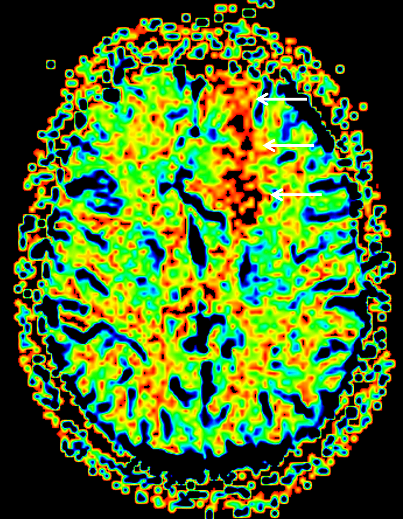
Cerebral blood volume map demonstrates relatively elevated cerebral blood volume corresponding to the left frontal mass (white arrows).
Diagnosis
Oligodendroglioma
Differential Diagnosis
Become a PRO member to unlock the differential diagnosis
Pearls
Become a PRO member to unlock the pearls