Findings
CT
- Peripherally-calcified mass along the medial right temporal lobe with adjacent small volume subarachnoid and subdural hemorrhage tracking along the floor of the right middle cranial fossa
MRI
- Giant aneurysm involving the right internal carotid artery terminus and M1 segment of the right MCA with saccular component measuring 2.7 x 1.2 x 1.7 cm
- Radiating pulsation artifact
- A more posterior fusiform component measuring up to 0.6 cm in thickness appears to supply right M2 branches, which remain patent
- Tiny (~0.2 cm) outpouching directed medially from the supraclinoid right internal carotid artery, possibly representing a tiny aneurysm or infundibulum associated with the superior hypophyseal artery
- Hypoplastic right A1 ACA segment
- Increased scattered subarachnoid hemorrhage, most abundant in the right sylvian fissure and sylvian cistern
- Small volume intraventricular hemorrhage layering in the occipital horns of the lateral ventricles
- Enlarged right cerebral convexity subdural hematoma measuring up to 5 mm in thickness
- No midline shift or evidence of herniation or hydrocephalus
Annotated Images & Illustrations
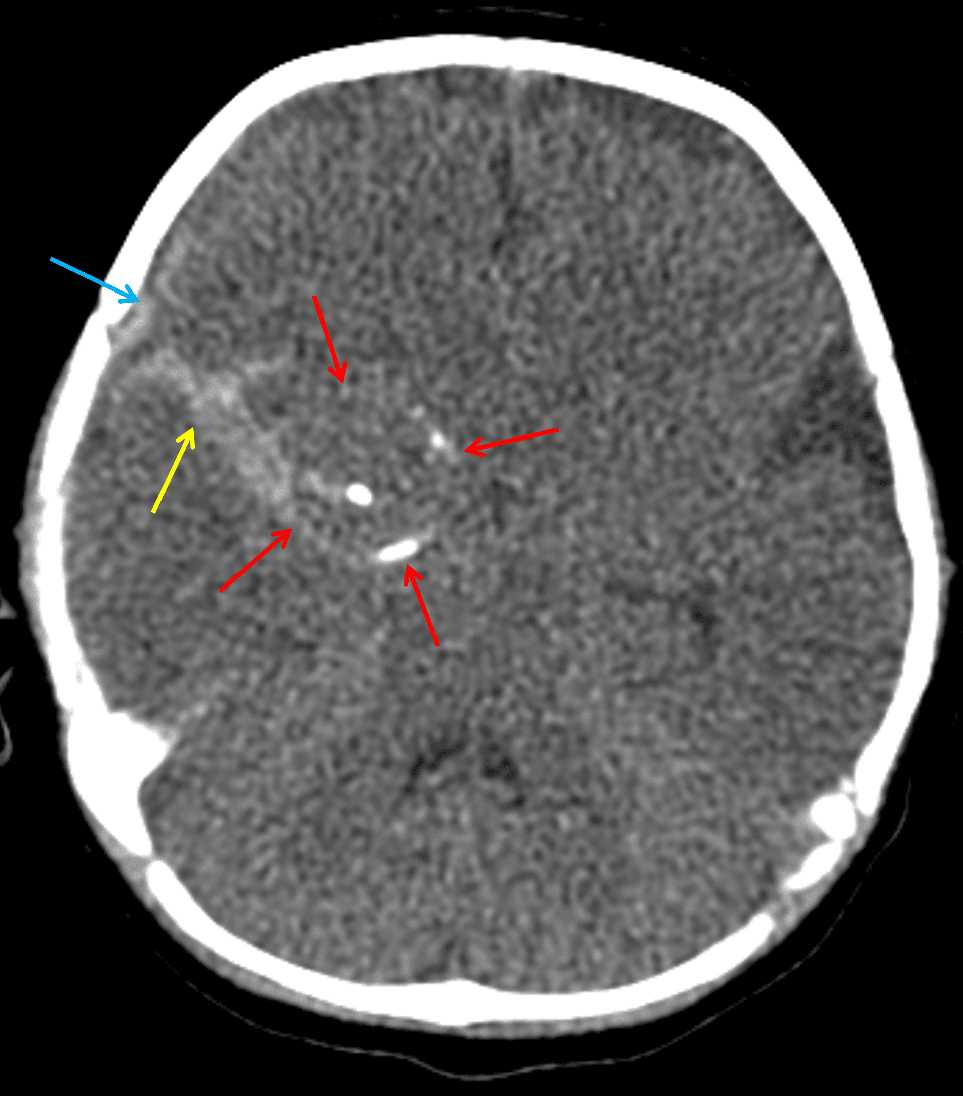
Peripherally-calcified lesion along the right temporal lobe (red arrows) with adjacent subarachnoid (yellow arrow) and subdural (blue arrow) hemorrhage.
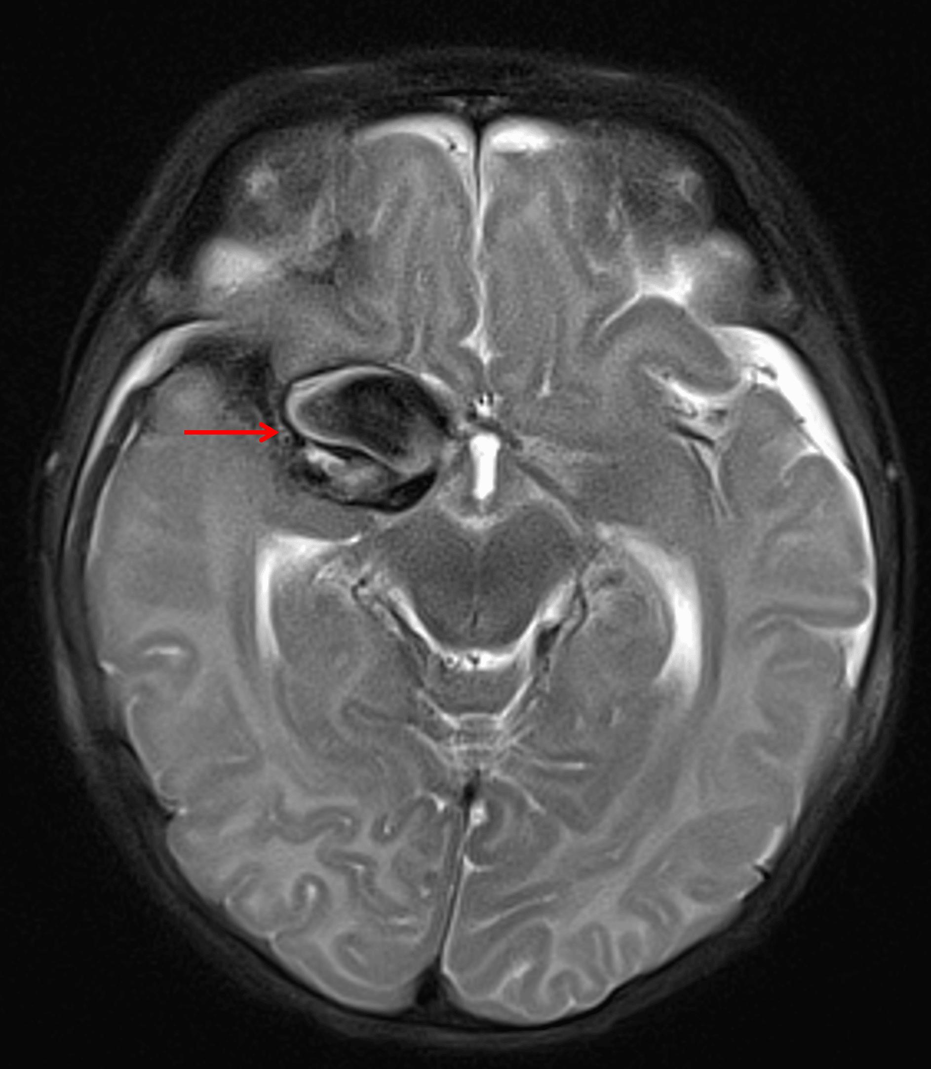
Corresponding markedly hypointense T2 signal with a swirling internal configuration (red arrow).
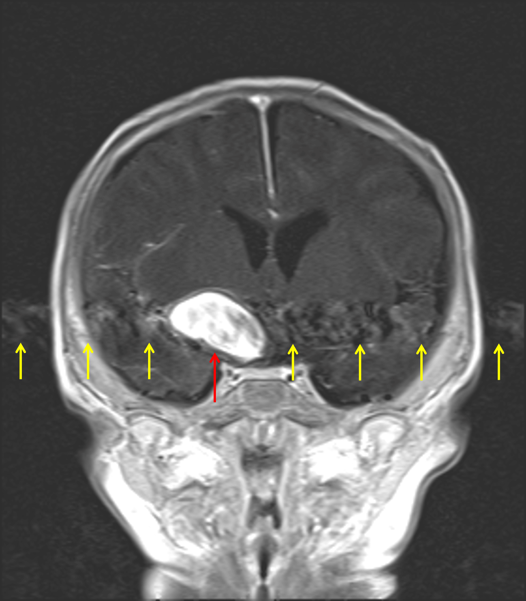
Diffuse internal enhancement on postcontrast imaging (red arrow) with corresponding radiating pulsation artifact (yellow arrows).
Diagnosis
Giant cerebral aneurysm
Key Imaging Features
Become a PRO member to unlock the key imaging features
Differential Diagnosis
Become a PRO member to unlock the differential diagnosis
Discussion
Pearls
Become a PRO member to unlock the pearls