Demographics:
6 years old, Male
Indication:
Seizure
Findings
CT
- Hyperattenuating lesion in the anterior right frontal lobe with a mixed attenuation mass with corresponding calcification along its lateral margin
- Surrounding vasogenic edema
MRI
- Irregular lesion in the anterior right frontal lobe with heterogeneous internal T2 signal intensity and a T2 hypointense rim, areas of intrinsic T1 signal hyperintensity, and extensive corresponding susceptibility artifact
- Corresponding areas of enhancement
- Adjacent circumscribed lesion measuring 4.5 x 3.5 x 3.3 cm with internal T2 signal hypointensity and a rim of susceptibility artifact, most consistent with a parenchymal hematoma
- Surrounding vasogenic edema and local mass effect resulting in slight right-to-left midline shift without evidence of downward herniation or hydrocephalus
Annotated Images & Illustrations
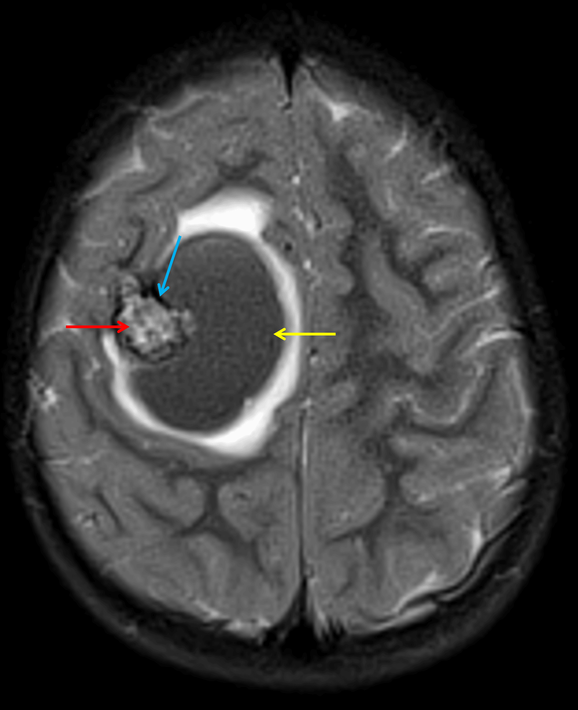
Lesion in the right frontal lobe with heterogeneous internal T2 signal intensity (red arrow), a rim of T2 signal hypointensity (blue arrow), and an adjacent hematoma (yellow arrow) with surrounding edema.
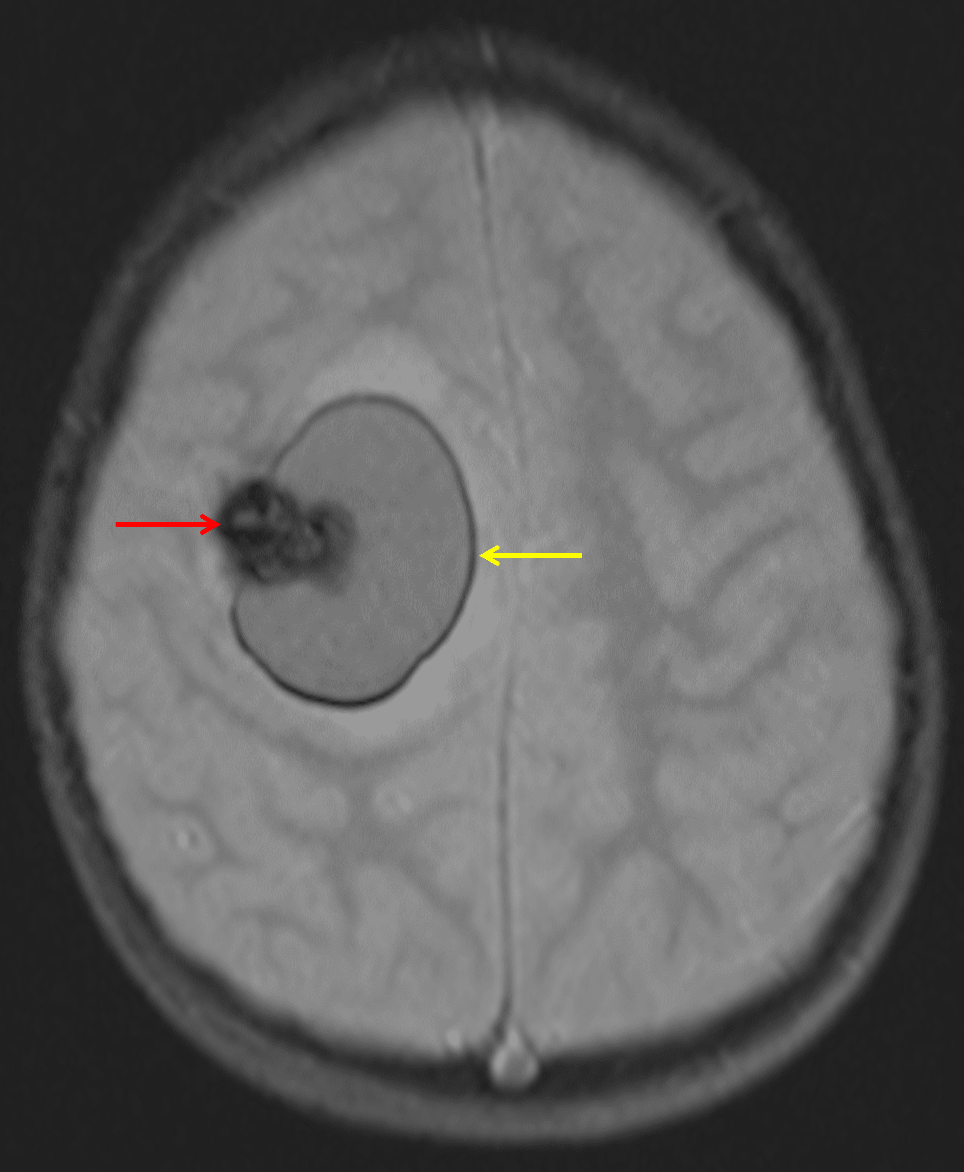
The lesion demonstrates extensive corresponding susceptibility artifact (red arrow). In addition, there is a rim of susceptibility artifact along the adjacent hematoma (yellow arrow).
Diagnosis
Cavernous malformation
Differential Diagnosis
Become a PRO member to unlock the differential diagnosis
Pearls
Become a PRO member to unlock the pearls