Demographics:
38 years old, Male
Indication:
Seizure
Case #1
Findings
- T1 hypointense, T2 hyperintense expansile mass extending to the cortex in the left superior frontal gyrus
- Mixed hyperintense and isointense internal FLAIR signal
- No corresponding enhancement, restricted diffusion, or susceptibility artifact
- Mild corresponding local mass effect without midline shift or evidence of herniation or hydrocephalus
Annotated Images & Illustrations
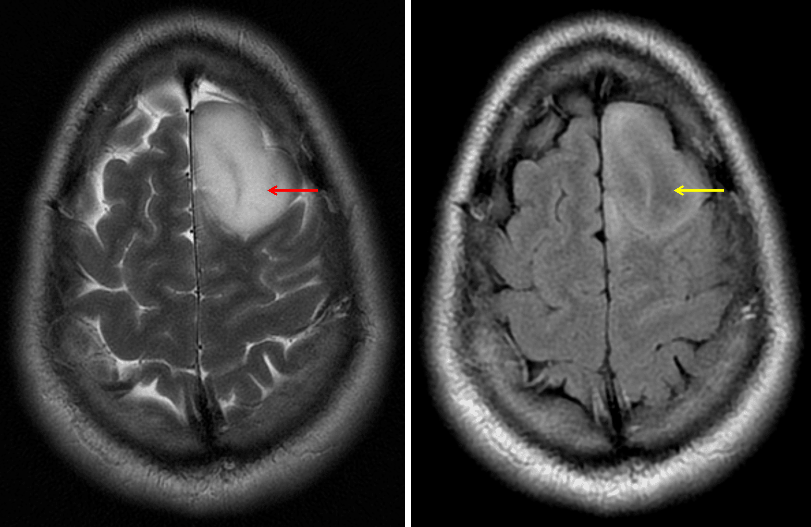
The T2-FLAIR mismatch sign: the lesion demonstrates internal T2 signal hyperintensity (red arrow) contrasted to relatively hypointense FLAIR signal (yellow arrow).
Diagnosis
IDH-mutant astrocytoma
Differential Diagnosis
Become a PRO member to unlock the differential diagnosis
Pearls
Become a PRO member to unlock the pearls
References