Findings
CT
- Areas of hypoattenuation in the anterior right basal ganglia/anterior limb of the right internal capsule and in the right frontal subcortical white matter with mild corresponding local mass effect
MRI
- Multifocal areas of parenchymal restricted diffusion involving the bilateral cerebral and cerebellar hemispheres
- The largest lesion is located in the anterior right basal ganglia/anterior limb of the right internal capsule, which measures 3 x 2.5 x 2.3 cm, demonstrates peripheral enhancement which is incomplete along its ependymal margin, and demonstrates central restricted diffusion
- Linear enhancement extends along the superior margin of the right lateral ventricle
- Many of these lesions demonstrate peripheral susceptibility artifact
- Surrounding vasogenic edema, particularly involving the right basal ganglia and right frontal subcortical lesions
- No substantial midline shift or evidence of herniation or hydrocephalus
Annotated Images & Illustrations
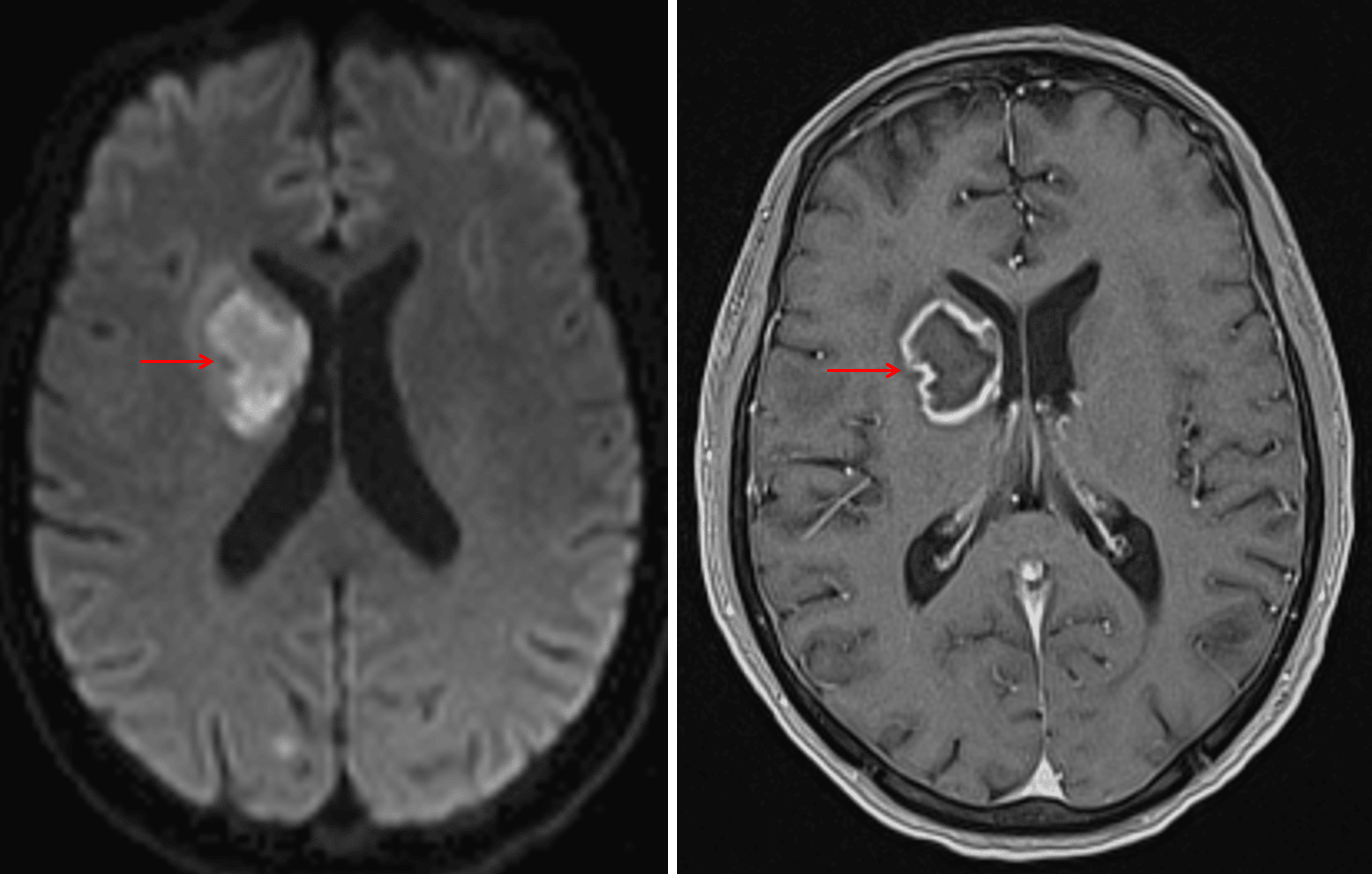
Peripherally-enhancing lesion with central restricted diffusion in the right basal ganglia and internal capsule (red arrows), highly concerning for a pyogenic abscess. Close proximity to the ventricular margin increases the risk for ventriculitis.
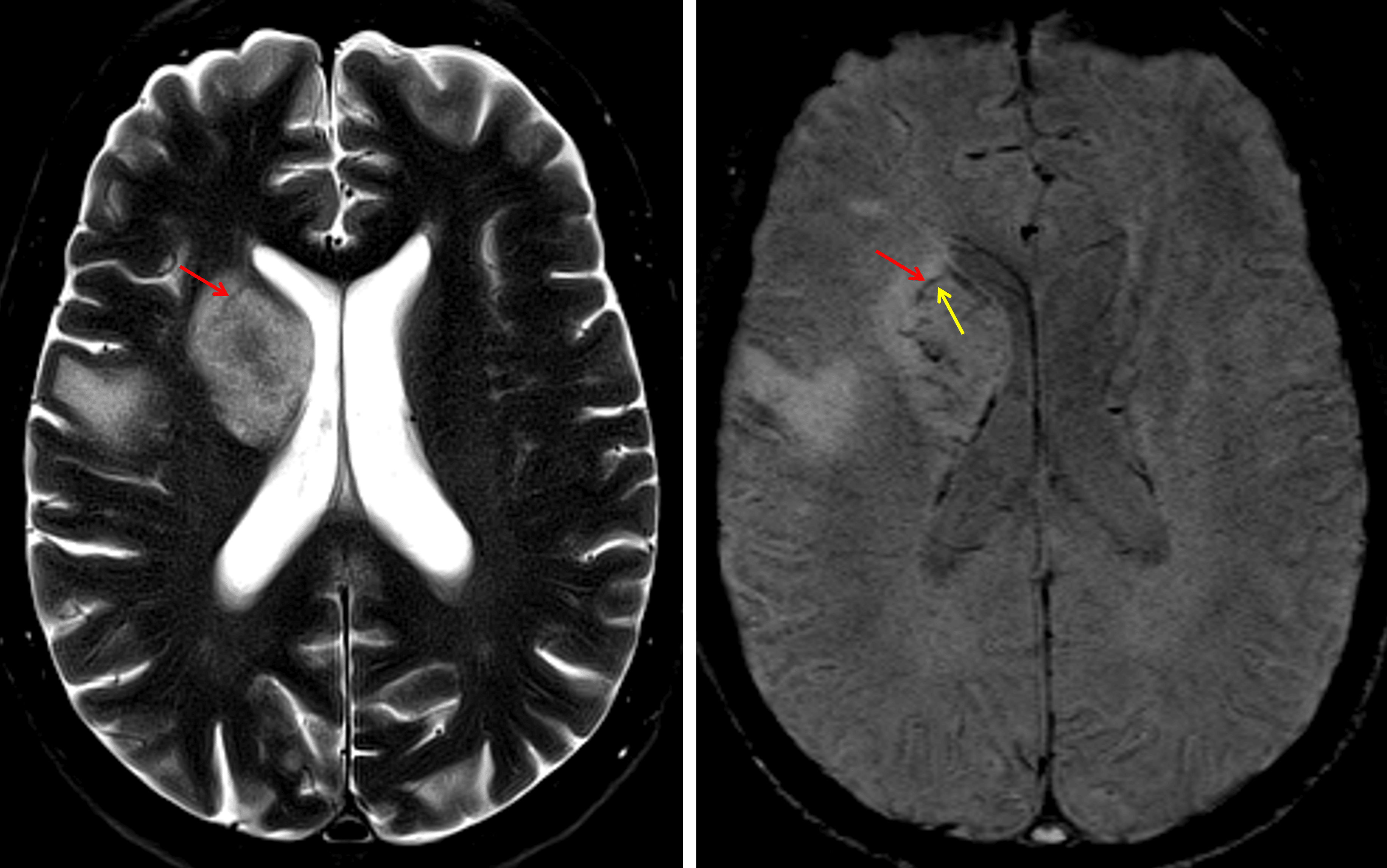
Although this case is not the best example of an abscess rim, there is a thin T2 and SWI hypointense rim (red arrows) with a thin internal SWI hyperintense ring (yellow arrow).
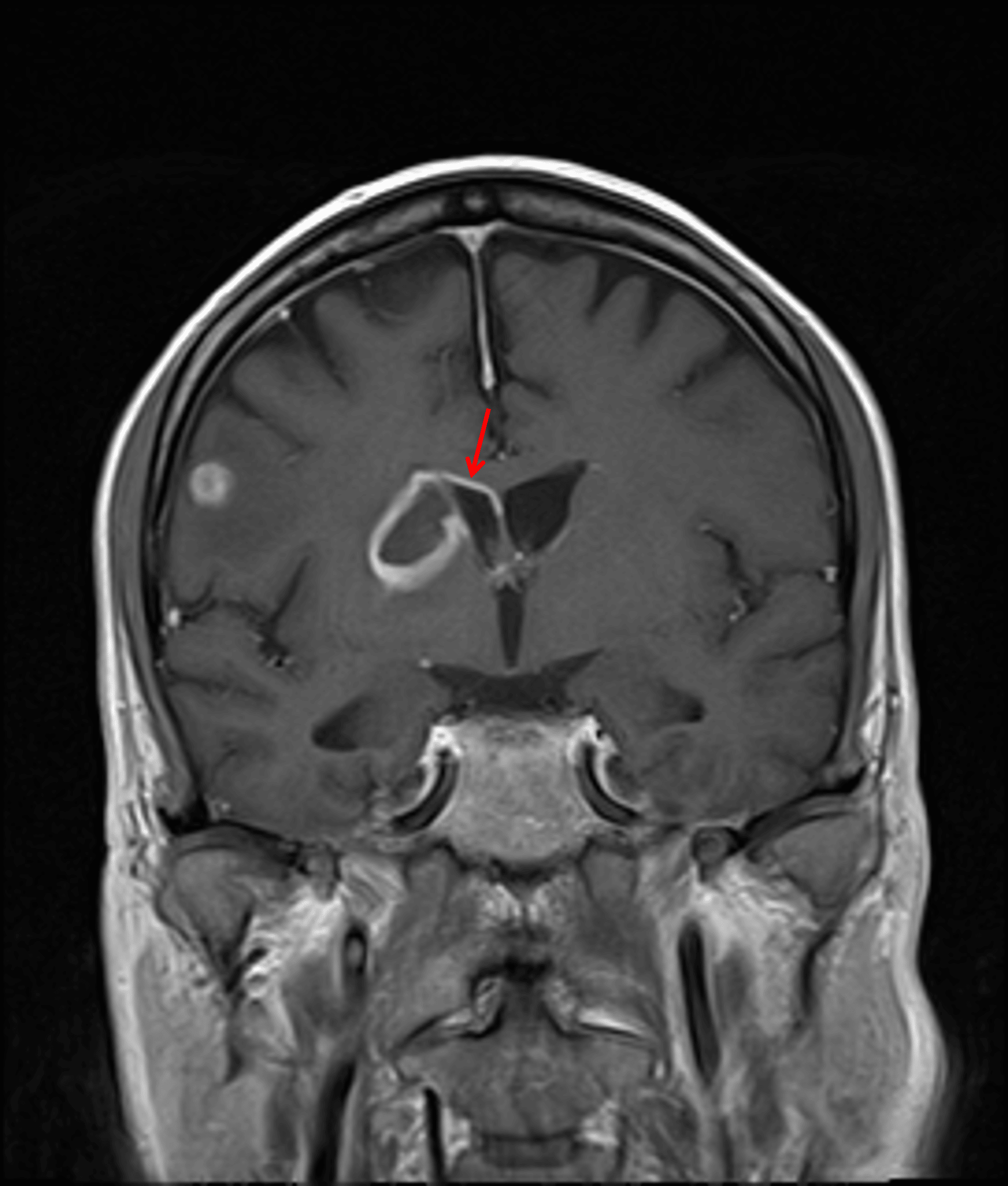
Enhancement extending along the superior margin of the right lateral ventricle (red arrow), which raises concern for developing ventriculitis.
Diagnosis
Septic emboli with pyogenic brain abscesses
Key Imaging Features
Become a PRO member to unlock the key imaging features
Differential Diagnosis
Become a PRO member to unlock the differential diagnosis
Discussion
Pearls
Become a PRO member to unlock the pearls